Tuesday, May 31, 2011

Throw away your 8-peak beads...now.

Why is it that each booth with a new piece of hardware I walked up to at CYTO had the same set of plots 'demonstrating' how 'sensitive' their instrument is?  I can't tell you how disheartening it is after years of clamoring for a re-definition of instrument 'sensitivity' to see marketing materials littered with histograms proudly displaying 8 peaks.  What does that actually mean?  and Why do instrument manufacturers design instruments around this 'standard'?
Now, I can't totally claim innocence here.  As you can see here, and here, I do use 8-peak beads as part of my panel of instrumentation tests.  However, it's not the only test I run, and I use it more so to dismiss potential problems than single out an instrument that is performing particularly well.  One of the best things 8-peaks can tell you about an instrument is the presence of background due to laser light bleed-through, or possibly a bad filter.  8-peaks are also pretty useful as an all-around alignment bead.  Beyond that, there's not much you can infer from the resolution of 8-peak beads as to the ability of said instrument to resolve dimly stained cells labeled with a particular fluorochrome from unstained cells.  For example, the ability to resolve 8-peaks in the FITC channel doesn't mean a whole lot as to its ability to resolve dim GFP signal from negative cells.  Likewise, the inability to resolve 8-peaks doesn't necessarily mean that channel will perform poorly for a dim fluorescence signal.  Let's look at an example.
I recently had the pleasure of evaluating the 8HT from EMD-Millipore (check out the full review here).  For comparison's sake, I ran the same set of tubes on our Beckman Coulter Gallios.  After collecting the data and doing the requisite comparisons, I noticed how closely the 8-peak data matched between the two, especially in the far red channel where we'd usually detect PECy7.  The first slide below shows the 8-peak data for the PECy7 channel.  The last 3 or 4 peaks are pretty much overlapping with one another.  If we were using the 8-peaks as a metric of 'sensitivity' we'd probably conclude that these two instruments are pretty similar in their ability to resolve dimly fluorescent PECy7 stained cells from unstained cells, right?...right?


Not so fast.  If we stain capture beads with a PECy7 antibody and look at the ability to resolve the 4 peaks that represent different levels of antigen density from each other as well as a blank bead, we can better assess (emphasis on the second syllable, please) the true 'sensitivity' of the two instruments.  Looking below at this figure we can easily see that the Gallios is able to resolve PECy7 much better than the 8HT.  This conclusion matches perfectly with real-world staining examples run on these instruments.  It's obvious that if PECy7 was a pretty darn important conjugate for my panels, you know which instrument I'd be buying (if I could afford it, that is).


So, what's the take-home message here?  Well, it's simple, 8-peak data cannot be used as a surrogate for how well an instrument will detect your panel of fluorochromes.  What should you do?  Again, simple:  make sure YOU run YOUR favorite flavors of fluors on the instrument you're evaluating to give you an idea of how well it's set up for YOUR experiments.  You can certainly do this by staining your cell samples of choice, or you could use a multi-peak capture bead to look at resolution.  And if you want to quantitate this a bit more, you could extrapolate the peaks down to an area just above the blank bead to determine precisely how dim of a population you'd be able to resolve.


Tuesday, May 17, 2011

EMD-Millipore 8HT Review

In my quest to find a mid-range cytometer to replace my ailing FACSCantos, I've come upon the 8HT from EMD-Millipore (whom I'll probably just call Millipore for now, or maybe even Guava at times). The 8HT is a 2 laser, 8-parameter cytometer (2 Scatter and 6 Fluorescence, setup in a 4-2 configuration).  It represents the "top-of-the-line" instrument in the field of 8 cytometers that vary in their number of lasers, detectors and the absence/presence of a microtiter plate loader.  The 8HT can accept either a 96 well plate or 10, 1.5mL microfuge tubes.  There are also a set of 6 tube slots for various washing/rinsing purposes.  There are a few unique features to this instrument which I'll briefly describe below before getting into the data.

All of the legacy Guava instruments and the new Millipore additions share the same microcapillary fluidics system.  Whereas in flow-cell containing instruments, a sheath fluid is funneled through a conical shaped flow-cell where it creates the hydrodynamic force used to align the sample core stream through the laser interrogation point, here there is no sheath fluid.  The microcapillary system is basically a clear straw through which the sample is drawn, and the microcapillary walls provide the physical barrier to align the sample, essentially replacing the sheath fluid.  This basically means there's no PBS tanks to fill, and no giant waste container to dump down the sink.  Also, since the sample core cannot expand in diameter to increase event rate like it would in a hydrodynamically focused system, the way in which you increase event rate is by literally increasing the speed at which the fluid is drawn through the capillary.  With the capillary system, you do get a bonus in absolute counts for everything.

A second unique feature of the system is the modulated laser setup they've implemented to get around the need for separate pinholes and laser delays.  Basically, the 488nm and 640nm laser lines are modulated out-of-phase with each other at a high frequency so that red fluorescence emitting from the cell while the blue laser is exciting is sensed separately from red fluorescence emitting from the cell while the red laser is exciting.  I was pretty skeptical at first, but this works surprisingly well.  For example, APC fluorescence was pretty well excluded from the Red1 channel (Blue excitation/Red emission).  What makes this remarkable is that Red1 and Red2 are actually the same detector, but because of the modulation, the emission is able to be separated out cleanly.  

The software, InCyte was pretty good.  The thing that bugs me a little is the mere presence of the old Guava modules.  The 8HT software, in general seems a bit schizophrenic.  You can jump back and forth between a green background Guava software to a grey background InCyte.  I think it would be less confusing if there were just one platform to use.  The InCyte software is pretty capable on its own, so I can't see what's the use of the Guava software.  I'm not one to use canned application-specific templates, so that makes anything with a green background pretty much useless.  There are a few unique analysis tricks built-in as well, which show your data in a heat map-like graphic.  However, probably my most favorite feature of the software is something fairly minor.  To adjust the threshold on FSC, you can simply drag a red dotted line up and down the scale.  There is no confusion on where the threshold is set.

So far, so good, right?  Well, here's where things fall apart - Data.  Like many of the other units I've tested in this range, their fluorescence resolution seems to be lacking.  I'd put the 8HT pretty much on-par with the rest, but let's take a look at some figures.  I ran my standard battery of tests including, Single Peak UltraRainbows (to get a glimpse at alignment via the CV), 8-peak rainbows (to assure a certain level of dynamic range and resolution), PI stained CENs (to look at linearity as well as well-to-well carryover), and my 'gold-standard' dim population resolution (using antibody stained capture beads).  This time, to mix things up a bit, I chose to run the EXACT same samples on our Gallios, and just to make things absolutely fair, the samples were run on the 8HT first and then on the Gallios (just in case they started to deteriorate over time).

Single peak URFP, showing CVs of the fluorescence channels on the 8HT versus the Gallios.


8HT URFP Bead CV
Gallios URFP Bead CV


Next up, the ubiquitous 8-peak Rainbow Beads.

8HT 8-peak Rainbow Bead Resolution

Gallios 8-peak Rainbow Bead Resolution
PI Stained CENs:  Here we notice a problem with the 8HT.  My guess is that the unbound PI in solution is too much for the system to handle, and the detector is being swamped by light.  Obviously, this could be alleviated by titrating the PI out, but as you can see below, the Gallios' baseline restoration has no problem with the unbound PI.  If you think about it, in a capillary system, the amount of sample fluid that is illuminated along with the cell is much higher than in a hydrodynamically focused system running at a narrow core stream, so unbound fluorochrome in solution surrounding the cells has a huge impact on background.  Keep this in mind when looking at the Dim population resolution data.  Also, for carryover, I'm simply looking for cells in the subsequent well to have been stained by PI carried-over from the prior well.  In this case, carryover appears to be less than 1:10,000.


For the Dim Population test, antibody binding beads stained with different fluorescently labelled CD4 antibodies were run on the 8HT at the 'very low' flow rate and the 'medium' flow rate.  There are suppose to be 4 stained peaks (blue) and an unstained peak (grey).  The lowest stained peak represents about 2500 bound antibodies (CD4 on Human PBMCs is about 50,000 binding sites).  Here, we get a really great picture of the background issues.  When looking at a blank bead by itself, the background is pretty low.  However, once you have some fluorescence present, the background peaks merge together and offer no resolution.  This effect is enhanced when you increase the flow rate.  Again, the exact same samples are run on the Gallios.


So, as I've said many times, the convenience that these systems offer may be nice, but I'm not sure it outweighs the lack of fluorescence resolution.  I'm sure things could be optimized so that this system would perform better, but I'll always come back to the fact that we don't run into these problems on our LSRII, Fortessa, Gallios, or even our Cantos, Caliburs and Scans.  What's the difference?  Hydrodyanmically focused streams with gel-coupled collection optics, high-quality/high-powered lasers, and bit-dense/fast sampling electronics.  The quest continues...

Monday, April 18, 2011

Display Transformation and FlowJo: Confused? Read on.

I'm not going to discuss the merits of transforming your log-scaled fluorescence flow cytometry data;  I'll leave that to the professionals.  What I will try to elucidate here is how I tweak the transformation using FlowJo (Mac version 9.3, sorry Windows people).  For simplicity, I will use the factory default preferences to start.  It's important to note that if you're on a shared computer, the preferences could have been changed, which may result in some funky things happening.  It's probably best to get a set of preferences that you like, and save them so each time you can pull up your preferences.  The preferences that matter for this are the Compensation section in the workspace tab (red box) and the "Define" button for  32-bit data, which will open the Options window for these type of data.
There are basically 3 things you can modify when adjusting the display transformation.  They are the Number of decades, Additional negative display size and the Width basis. The Number of decades controls, to a large extent, how many decades of dynamic range is shown for events greater than zero. By default, this is 4.5--even if you export 5.5 decade data, use 4-4.5, otherwise too much visual space is devoted to the lowest decade. Additional negative display size controls, to a large extent, how much visual space to devote to events that have values less than zero. Since we are displaying data on a log scale, zero is not defined. So, there needs to be a way to display data around zero that makes sense. This is what's at the heart of the biexponential display. Near zero, the log scale becomes linear so that zero can be defined, and then goes back to log when safely in the negative realm. The number of channels around zero that are transformed into the linear realm is defined by the width basis. By default, FACSDiVa uses a width basis of -100, whereas FlowJo's default is -10.

So, how does this affect your data?  Well let's take a look at an example.  FITC and PE stained capture beads were run on a FACSCanto and exported in the FCS 3.0 format.  No compensation was done at the instrument, and single stained controls were used to compensate the data in FlowJo.  Below is the uncompensated data file as well as a compensated file using the defaults that were currently applied.  

Looks pretty good, but let's take a look at some simple tweaks.  For this, we'll go to the Platform menu, then Biexponential Transformation and then Manually Specify Transformation, which will bring open the window to edit the transformation settings.

Again, we have the option to change the width basis, Positive decades and Additional Negative Decades.  Let's assume we're not going to change the Positive decades, so we'll focus on what the width basis and negative decades will make.  Below are the plots shown at each of the width basis presets.  As you can see, the main affect is squishing the data closer together around the zero point.  Good to get data off the axis, but you can easily take it too far and end up reducing your ability to resolve dimly stained cells from unstained cells.

The question then is how much transformation do you apply?  One strategy that I like to employ is to try and visualize this better with a contour.  The goal is to remove the bimodal-like profile of the populations as they cross the zero point.  Once I'm able to do that, I then increase the amount of negative log space so that most of the data is not on the axis.  For example, below I show a -10 width basis, 1 additional negative log in a contour plot.  In the brightest peaks, it is easy to see a pronounced dumbbell shaped population straddling the zero point.  If I modify the width basis a bit to get rid of the dumbbell shape, and then reduce the additional negative space to remove extraneous white space, I get a profile like the one on the right.  Notice that each axis is done separately and can have different width basis and negative logs to achieve the best transformation.  


Once all is said and done, I now have a well transformed plot that is worthy of publication.  Below is the original uncompensated plot, the default transformation, and the modified transformation.


A few tweaks and a bit of trial and error is all you need to get visually pleasing plots that will actually help you make better decisions in terms of region drawing and data interpretation.  So, please feel free to play around with these settings and see how well you can transform.


Thursday, March 31, 2011

Sort Schedule Changes: The Who, What and Why.

Let's start at the beginning.  Back in the days of the FACStar Plus and water cooled gas lasers, we would begin accepting sort reservations at 10AM.  This allowed us to get in at 9AM, warm up the lasers for 30 minutes, get the fluidics going, and have everything running smoothly by 10AM.  We typically did 1 or (on a good day) 2 sorts, which lasted 2-4 hours going at a rate of about 5000 events per second.  Adding the MoFlo in 2001 allowed us greater throughput by allowing us to run at 10's of thousands of cells per second, but we still needed a good amount of time to get started in the morning so the I-90s could warm up and the fluidics could stabilize.

As word got out about sorting applications in general, the sorters became busier and busier; so much so, that we traded in the old FACStar Plus, and traded up to the FACSAria.  Around the same time, we retired our water cooled gas lasers, and outfitted the MoFlo with the solid state Lyt-200 from iCyt.  The ease of setup on the Aria, and the nearly instant-on of the solid state lasers made getting started in the morning much easier.  Slowly but surely, we allowed earlier and earlier sorts until we officially added the 9AM - 10AM slot on both sorters.  The increased capacity kept up with demand for a period of time, but soon even running both sorters 9AM - 5PM wasn't enough.  We also saw that the afternoon slots were filled weeks in advance, and the unfortunate few who had to book sorts last minute were stuck with the 9AM slot, usually forcing them to get started in the wee hours of the morning.  In an attempt to solve this problem, and position ourselves for an S-10 application for a 3rd sorter, we added a 5PM - 8PM "late night" sorting slot on Tuesdays and Thursdays, and had one of the operators work a 12PM - 8PM shift on those days.  These slots were gobbled up instantly, and soon became a favored slot amongst the users.  Still struggling with overall capacity, the cancer center stepped up and helped us purchase another sorter, the FACSAria II.  So, now we had 3 sorters available from 9AM - 5PM Monday thru Friday, and the FACSAria II available an additional 3 hours on Tuesday and Thursday evening.  This setup served us very well for a couple of years.  Along the way, we still saw that afternoons filled up very quickly, and the 9AM slot was still being used for last minute sorts, or GFP cell line sorts.

So, we decided to take a look at usage more closely, and query our user group about the possibility of opening more 5PM - 8PM slots.  Of course everyone thought the idea of more evening slots was great, but we needed some data to support this change.  The figure below shows the frequency in a year of a sorter being used at each hour of the day.  It is separated out for each sorter.  The blue line shows what percentage of available time the sorters (as a whole) are being used.  As expected, the afternoon slots are used 70% of the time, while the 9AM slot is reserved only 25% of the time.  Also, the 5PM - 7PM slots are used 60% of the time, even though the actual number of sorts taking place appears low.  The 25% number at 9AM is also a bit misleading.  This represents that hour being booked, but I can tell you, it is very rare for someone who books a sort from 9AM until 11AM to actually show up at 9AM, and the longer that reservation is, the later they show up.  So, the actual usage of the 9AM hour is probably much lower.  I'd estimate it at 10% or so.  So, instead of sitting around waiting for the 9AM sort to show up, we could utilize that time better by getting QC done on our army of bench-top analyzers, or allow for more stabilization time of the sorters to get even better results.  So, beginning in April, we will have new sorting hours as follows:  M - F 10AM -5PM, and additionally on one sorter, 5PM - 8PM M - Th (no one wants to sort until 8PM on Friday night!).  The number of available hours per week is still about the same.  Previously, we offered 121 hours of capacity each week, and now we're offering 117 hours. Our actual weekly usage is about 70% of that anyhow, so we feel the new hours are plenty for the current demand.  In addition to all this, we have been aggressively pursuing users who are frequent FACSAria users to get trained and run their own sorts.  We have half a dozen users who already do this on a regular basis and will be tapping a few more competent users in the near future.  We feel this, along with the increased evening hours, and the removal of the 9AM slot, will maximize the efficiency of our sorters and allow more convenient access for our users.

Wednesday, March 23, 2011

Getting in tune with the Attune

An updated review of the Attune Cytometer, dated October 2012 is now available:  You can reach that post here:  http://ucflow.blogspot.com/2012/10/life-technologies-attune-cytometer-deja.html

EDIT 2:  I've met with some folks from Life Technologies and they feel the instrument that was set up in my lab for demo was not properly aligned, and so I've agreed to give it another try.  I'm not sure if I'll use the Violet/Blue combo or the Red/Blue system, but either way, I'll post the results and reference back to this post when I have them.

EDIT:  Regarding the statement below in the Optics section, "I have not received information that I requested yet, but the 'PMTs' do not appear to be 'PMTs'."  I have heard back and there are, in fact, PMTs in the system.  It's just not obviously visible when you simply open the hood.  They are the Hamamatsu H10720 Series, and in the far red channel, they are using the Red sensitive "-20's"

You've probably heard about the novel approach to sample focusing on the Attune, and you've probably even read through some of the marketing materials distributed by Applied Biosystems (Life Technologies).  So I won't go through all those features in detail, but I will share my impressions and tell you what it means for your data in the end.

Briefly, the Attune is a 2-laser, 6-fluorescence detector small-footprint cytometer.  The fluidics are syringe pump driven (as opposed to pressure-driven), and it's in the focusing of the cells through the laser interrogation point that is unique.  Instead of hydrodynamic focusing applied by a sheath fluid (such as PBS), the Attune uses an acoustic wave form to push the cells into the center of the stream, forcing them to line up in single file.  One advantage of this is the small amount of sheath buffer you need to put into the system (roughly 1L per day compared to 6-8L for a hydrodynamically focused system).  Another benefit is the ability to push through large quantities of sample fluid per unit time.  Whereas a pressurized, hydrodynamically focused system might top out at about 200ul/minute, the Attune can achieve volume flow rates of 1000ul/minute.  It can also maintain the CV of the fluorescence signals even at high flow rates. Lastly, the system can actually change how fast the events are passing through the laser interrogation point.  They have a standard, and high-sensitivity setting, which tells the system to pass them through at the normal rate, or at a slower rate, respectively.  The idea is that if the cells pass through the lasers more slowly, the fluorochromes will emit more photons, allowing you to detect lower amounts of fluorescence.

Optically, the system has a 20mW 488nm laser and a 50mW 405nm laser.  There are no other options for laser lines at this point.  3 of the fluorescence detectors are dedicated to the blue laser and 3 to the violet laser, and they're set up for FITC, PE, PerCP/PECy7, and PacBlue, Qdot 525, and Qdot 605/PacOrange.  The filters are easily changeable, and there's even a storage section for spare filters (how nice!).

I'll try to break up my comments by instrument components.  I will say, many of the complaints I had regarding the software were said to be fixed in an upcoming release, but I will give my opinions based on the instrument I evaluated (03/09/11).  Also note that the instrument was installed by a field service engineer and passed their performance and tracking protocol (similar to CS&T a la BD).

Fluidics:
1.  Being a syringe pump system, you need to specify how much volume you wish to analyze before you even put on  your sample.  Of course, you can always add to your sample acquisition, but it doesn't have the 'free-run' capabilities that a pressurized system has.
2.  If you tell it to take up 500ul of sample, and then you get enough data, or you want to just take your tube off, the remaining sample in the line goes to waste, and not back to your tube.  So you could foresee a possible loss of sample in some situations.
3.  The dead volume is huge!  200ul of your sample is taken regardless of how much sample you wish to analyze.  So, if you want to analyze 100ul, you need to have at least 300ul of volume in your sample.
4.  It was really nice being able to put on different sizes of tubes and not have to worry if they're the right kind or cracked.
5.  As advertised, the CV's are really low even going at high sample throughput rates.
6.  Although they claim they can run at rates of up to 20,000 events per second, there is no report of the coincident rate going at that speed.  A high abort rate would obviously have an impact on detecting and counting rare events.  Even on our systems with zero "dead time" we still see coincident events to some degree.
7.  Although in theory we'd expect the high sensitivity setting (i.e. slowing the cells down in the interrogation point) to increase resolution, my tests did not show any improvement with stained beads (data not shown).  It's likely that the components that contribute to autofluorescence also increase their intensity so the separation between positive and negative doesn't really change.
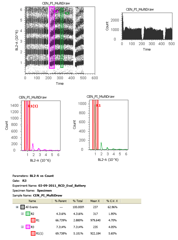
8.  When you tell the system to collect 2mL of sample volume, it does so in (for example) 500ul 'draws' from the tube, i.e. 4 draws from the sample tube.  The reason is that they don't want to have all that volume sitting in the tubing waiting to go to the flow cell.  The result is that you end up having these pauses in acquisition while the system refills the tubing.  Because of this, when you start collecting your sample again, the core stream has not completely stabilized leading to a decrease in fluorescence and a widening of the CV (Data Below).  In order to get rid of this data, which will surely mess up your analysis, you'd have to go to each point where it drew more sample and exclude the first few seconds of collection until the stream has stabilized.  It's an easy enough fix to make the software ignore data being collected for a few seconds every time it draws, and hopefully they'll implement that fix.

Software:
1. IT'S FREE!!!!!!!!
2.  The overall look of the software steals a page from Microsoft Office, with a prominent ribbon at the top for common tasks, and lots of right-click functionality.  Users should feel comfortable here.
3.  Similar to FACSDiVa software, there is a Experiment Explorer (Browser) that displays your experiment, all the tubes in the experiment, and the settings/plots saved within.  There was some confusion in terms of things being applied globally (to the entire experiment) or to an individual tube. Again, similar to DiVa, it's not always obvious if something is applied globally/individually, and how to change that status easily.  Lastly, it also resembled the Gallios software in that if you made a change to the plots/regions, you had to click save or else you'd lose those changes when you switched tubes.
4.  Gating was somewhat cumbersome.  Changing hierarchy of the gating structure after you've created gates was difficult and not intuitive.
5.  Displaying large numbers of events was slow and jerky.  There was this weird need to press 'F5' to refresh plots with lots of data point.

Optics:
1.  I have not received information that I requested yet, but the 'PMTs' do not appear to be 'PMTs'.  There is definitely not a vacuum tube with anode, dynodes and cathode like you'd expect to see.   It looks like some sort of silicon diode, but like I said, I haven't heard yet.  This could contribute somewhat to the performance I observed.
2.  I really appreciate the extra slots for filters on the instrument.  I'm a big believer in spare filters, but I can never seem to keep track of them (just look in our MoFlo room).
3.  Although the laser powers seem appropriate, there was no mention of how much of that light actually makes it to the flow cell.

Electronics:
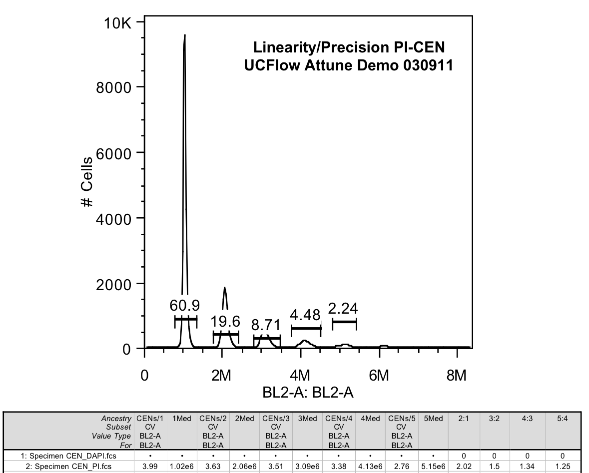
1.  Testing of linearity across the 6-log scale was good (CEN data below)
2.  23-bit ADC conversion yield 8.4million channels across 6 decades, which generates approximately 76 bins in the 1st decade.  This seems pretty good, and would allow for resolution of closely related populations on the low end of the scale, but that's not really what I saw.  I was able to see some 'picket fencing' which tells me that the noise of the ADC makes the bit conversion probably closer to 20 bit, which yield about 9 channels of resolution in the first decade.  This is speculation on my part since I do not know (yet) what ADC they're using, and what the noise level of that ADC is.

Data:  For this I ran my standard battery of tests, the results of which can be found below.

8-peaks:  Testing the overall alignment and diagnose background problems due to laser light.
Assessment:  8 peak resolution off the blue laser was not as good as other 'full-size' cytometers (i.e. FACSCanto-II, or Gallios)

CEN Linearity:  Using PI stained (and DAPI stained, but not shown) CENs.
Assessment:  Linearity was good along all of the scale tested.
Dim Population Resolution:  Here we have Antibody binding beads from Bangs stained with a FITC, PE or PacBlue antibody to demonstrate the ability to resolve increasingly dimmer fluorescence from background.  The more separated the peaks, and the further from the blank the better the resolution.
Assessment:  The FITC channel (BL1) seemed to be the worst of the 3 tested.  The PE channel was ok, and the PacBlue Channel (VL1) was pretty good.  To see comparable data on another instrument, check out the same data on the FACSCanto-II


Stream Stability:  For this, I tried to have it draw sample multiple times while looking at PI fluorescence in linear.
Assessment:  As you can see, the fluorescence dips down every time this fluidics draws more sample.  This leads to an overall higher CV if you don't time gate the data.  You can easily see this by comparing the purple box at the beginning of acquisition post draw and the green box that's mid-way through that collection.  

Final Thoughts:  Like many of the smaller sized cytometers, the Attune will probably handle most of the general types of applications, but will not compare to high-end cytometers in its ability to detect dimly stained populations, even using its High Sensitivity mode.  The real advantage is its ability to push large amounts of volume of a relatively dilute sample through the instrument without increasing CVs of the populations.  For example, a lyse/no-wash rare event blood tube.  In most every other case, we would simply concentrate the sample by spinning it down, and run it in a similar amount of time as what can be achieved on the Attune.  The overall look and feel of the software is fairly polished.  If they're able to correct a few major things (like return unused sample, not store data during stream restabilization, allow for a quicker refresh of data on the screen, and get rid of the F5 necessity) with some software changes, it will be a much better instrument.  The lack of a red laser, or laser options is a real hurdle for many people, especially those with a large stock of APC and APCCy7 antibodies, and could be a deal-breaker for many labs.  It would also be nice to have a higher powered 488nm option as well.  This, along with the whole 'PMT' issue might lead to better resolution in the FITC channel, which was especially poor.


Thursday, March 10, 2011

Just when I thought I was out...they pull me back in.

I'm speaking, of course, about BD and the FACSCanto platform. Now, if you know me and have talked to me about flow cytometers, you know I haven't been too kind to BD and the FACSCanto-A. We have, and continue to, battled with problems on these instruments. It doesn't help much that we have hundreds of users running all sorts of who-knows-what through the instruments. But then again, our LSRII's never break-down, our 15 year-old FACScan and 10 year-old FACSCalibur never break on us and they get just as much use. Recently, however, I happen to have the privilege of running a FACSCanto II with an HTS. We've had it in the lab for a few weeks, so I've been playing around with it trying my darndest to break it, without success. It's definitely not a fair comparison as far as the use and abuse our other cytometers deal with, but I've tried to run some unfiltered chunky samples on it a few times, and it recovers well. The HTS has been running as well as could be expected; so well, that we're now putting one on our Fortessa. There are two other things on the Canto-II that have really caught my attention. The first thing is the overall fluorescence sensitivity and resolution. It certainly ranks among the top instruments I've ever tested. I ran my standard battery of tests on the instrument (dim population resolution, linearity, dynamic range, and precision) and the FACSCanto-II excelled in all respects. I'll highlight a few of these tests with some figures below. Before that, I want to document just how awesome it is to be able to put on a regular 12x75 tube with about 100ul, and be able to analyze almost all 100ul out of the tube. The way that the lever pushes the tube all the way up so that the probe is nearly hitting the bottom is awesome. That, combined with the absence of a DCM sleeve on the SIT makes this possible.  Dead volume is about as minimal as can be for a tube loader.

Figure 1 below simply shows some antibody binding beads from Bangs stained with a CD4 antibody coupled to either FITC, PE, or eFluor450. The lowest stained peak represents a binding capacity of ~3000 antigen. To put that into perspective, CD4 on human Tcells is expressed at about 50,000 antigen. So, you can see that something with 10-fold less antigen density can easily be separated from background.


Figure 2 is simply displaying 8-peak bead data.


And Figure 3 is showing PI stained CEN's to show precision (% CV of the 1st peak) and linearity peak-to-peak ratios.

Monday, February 7, 2011

A 19.2V Drill with every sorter purchase?

As you've probably heard, Beckman Coulter put itself up for sale last year, and now it looks like they will be purchased by Washington based Danaher Corporation. Danaher is a conglomerate that owns the Craftsman hand tool brand as well as businesses in electronic testing equipment, dental equipment, and monitoring products. They also own the microscopy company Leica. What does this mean for the flow cytometry world? Probably not much, but the extra capital available from Danaher could only help R&D for flow cytometry, right? Time will tell. It seems like the MoFlo just can't find a stable home. Ever since Cytomation was acquired by Dako, the MoFlo has been passed around like a plate of crudités at a 6 year old's birthday party. My advice to Danaher and those making the transition from Beckman Coulter - forget everything you think you know about flow cytometry, come talk to the folks in trenches, and design a new instrument that's more than a "me-too" product. Good luck! Here's Beckman-Coulter's statement. And here's what Danaher has to say.