Tuesday, June 5, 2012

Fluorish vs. Chromocyte... READY, FIGHT!


One of the more difficult tasks people encounter when they start making multicolor flow cytometry panels is the process of finding antibodies, and knowing which fluorochromes to use.  There's the primary task of finding the specific antibodies you want to use in a few fluorochrome options (hopefully) and figuring out which ones would work best on the instruments available.  And then, there's a secondary task of optimizing the fluorochrome with the antibody in line with the adage:  Dim fluors on abundant antigen, bright flours on sparse antigen.  This secondary, but equally important task is a pretty advanced skill that is difficult to automate or build into an algorithm, although some companies, like Woodside Logic's CytoGenie, do a decent job of trying to handle this.  However, in order to get to this second step, we need to first aggregate all the information from a lot of sources as to what's available.  This step can be built into a utility, and it is precisely this step which I will discuss below.  In particular, I'm going to focus on two offerings that have come online in recent years.  One is the utility built into the Chromocyte web portal** under its 'calculate' tab, and the other is a utility developed by the parent of FlowJo called Fluorish.

After playing around with both services for a bit, I'll highlight a few areas which can serve as a good comparison of the two.  The areas I'll focus on are Catalog, Hardware Integration, and Usability.

Catalog:  First off, let's look at the catalog of antibodies available from the two sites.  As expected, there's a lot of overlap, and they both have the catalogs of all the major vendors you'd expect to see.  There were, however a few exceptions.  For example, Fluorish.com doesn't have the catalog of EMD-Millipore, GeneTex, Cytognos, antibodies-online, Gen-probe Diaclone, and IMGENEX Corp - all of whom appear on the Chromocyte site.  The Chromocyte site didn't have Miltenyi Biotec, and Sony/iCyt, both of which are present on Fluorish.com.  Whether this translates into a use-case where you're unable to find a specific antibody you're looking for or not is debatable.  I'd assume that nearly every antibody commercially available is likely to be on both sites, and if there is something not available, both sites prominently display a way to let them know so they can add it.  However, it should be noted that the Chromocyte list seems to be outrageously exhaustive; allowing you to select antibody species from Alligator to Zebrafish, and everything in between.  Another nice feature of both sites is the ability to select viability or other dyes, and fluorescent proteins so you can exclude those channels from the potential list of available fluorochromes.  So far, we'll call this a truce, but wait...there's more.  Fluorish.com, not only allows you to query all these commercial sources, but you can also add your own private stash to the database and allow those custom sources to be displayed in-line with the commercial sources.  It just so happens that I'm part of a core facility that also makes and conjugates antibodies, so this is a BIG DEAL for me.  Catalog Winner:  FLUORISH.COM 

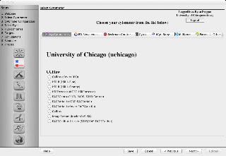
Fluorish's Panel Wizard showing Instruments in UCFlow Core.
Hardware Integration:  Next, I'm looking for the ability to integrate information regarding your facility's hardware.  Again, both sites offer the opportunity to input and save the configuration of your specific instrument.  Chromocyte uses a method where you select pre-defined filters from a list, whereas Fluorish has you input the cut-on and cut-off wavelength for each filter.  Obviously, if your specific filter is not in the Chromocyte list, you're out of luck (although you could request your filter to be added).  Using the Fluorish method allows you to precisely input your specific/custom filter, but it takes a bit of mental rigor (at least for my brain) as you try to calculate the cut-on and cut-off of a 595/14 BP filter.  Fluorish, however, takes things one step further by allowing you to export your FACSDiVa configuration file from CS&T and drop it in, yielding a hands-free hardware configuration process.  Both services have built in a layer of groups, which basically means I am set up on their site as a core facility, and other users can be part of my group, which gives them access to all the configurations I've already set up.  As you can see, both utilities have done a very nice job allowing you to build panels specific to your hardware configuration, however with the abundance of FACSDiVa systems, the extra ability to drag and drop instrument configurations from CS&T into Fluorish puts it just over top of Chromocyte.  Hardware Integration Winner (by a hair):  FLUORISH.COM

Usability:  Lastly, but most importantly is Usability.  This is such a huge topic, and one that's likely very personal to the individual user, that I'll simply report some of the things I really like or dislike about each of the services.  When it comes time to pick which antibodies you will use, I really like the interface of Chromocyte where they lay everything out in a grid pattern.  You can easily get a sense, at-a-glance, of how things might play out.  If there are only a couple options under one of the columns, you'll know you should probably take care of that column (i.e. channel) first.  The difficult part is that when you have a bunch of antibodies you're searching for and you take care of one of them (e.g. CD45), you'd expect the CD45 options in all the other channels would be greyed out or disappear.  However, it allows you to accidentally select CD45 in multiple colors.  Also, since the lists can sometimes be long, and if you choose one of the options that are off screen (i.e. an option you needed to scroll down to select), it would be nice if the column was greyed out or somehow highlighted to show you that channel is taken care of.
Chromocyte's interface for picking conjugated antibodies
Fluorish's answer to these problems is that it dims both antibody and fluorochrome options that remain once you choose your preference.  Although Fluorish's interface isn't as pretty as Chromocytes, it's definitely more functional and easier to use.

A nice feature of Fluorish that's missing from Chromocyte is the ability to specify a specific clone of an antibody.  So, if I know that I want to use RPA-T4 instead of OKT4, I can select that specific clone to refine my search.  

An advantage of Chromocyte is the fact that it lives completely in the cloud.  There's no software to download or upgrade, and so you're always using the latest version.  With Fluorish, there are two sides of the equation.  The first is Fluorish.com, which has limited functionality, but controls things like accounts, and core facility membership, etc...  The second part is a program that you download called the Panel Wizard (currently on beta version 1.0b34).  It works well, but completely fails at one crucial part of the equation... Mobile.  Now Chromocyte is by no means optimized for mobile, but it at least works.  Mobile is such a huge segment of my daily workflow, it's tough to forgive this omission on Fluorish's part.  However, it's desktop application does work quite well, and with it's nice usability features discussed above, it more than makes up for that omission.  Usability Winner:  FLUORISH.COM  

So, there you have it.  In terms of a free utility to help you build panels for your multicolor flow cytometry needs, Fluorish.com appears to be a better option.  **Of course, it must be said that Chromocyte has A LOT more to offer than its 'Calculate' tab.  With its wide breadth of resources on flow cytometry techniques, services, news, and tutorials, Chromocyte is a one-stop-shop for the flow cytometry community.  They've even plugged one of my blog posts, so you know they have good taste!  Now we just need Fluorish and Chromocyte to get together and make a baby so we can have to best of both worlds. 

Tuesday, March 27, 2012

7 Habits of a Highly Successful Core Facility

I know, I know, another spin on the "7 Habits..." meme, but hey, reserve your judgement until you've read through them.  This post is definitely geared towards core facilities, but I think you'll find this transcends to multiple service oriented businesses.  Let me know if your facility is currently implementing any of these strategies, or ones that I've missed.  And so, without further ado, I present the 7 Habits of Highly Successful Core Facilities, which was compiled after an exhaustive study of core facilities at the University of Chicago with an n of exactly 1 (i.e. us).

1.  Interact with OEMs - I cannot stress enough the importance of having regular conversations with those companies who are making the products you are using.  Many times labs will wait for a company to contact them with the latest and greatest products that have recently launched, however we've tried to take a proactive approach when it comes to new products and services.  This invariably leads to demonstrations or evaluations, or even early access to new tech as a beta tester.  In addition, you might just be able to voice your opinions so that future iterations of a product better suit your needs.

2.  Engage with your users regarding their scientific questions - I've said multiple times before, people love to talk about their projects, so why not give them another opportunity to fine tune their story.  I mean, you don't have anything better to do while sitting at the sorter, do you?  The flip side of this is you may actually pick up on something and be able to suggest a technical improvement of the assay.  There have been numerous times when someone is explaining to me what they're trying to do, and I've suggested using a different technology or approach, and it has worked out really well for them.  For example, a user who I was training on a bench-top analyzer was complaining that it's impossible to differentiate between necrotic cells and late apoptotic cells definitely by flow cytometry.  I simply suggested using our ImageStreamX instead (which also measures morphological parameters such as area of cytoplasm and darkfield scatter - a characteristic of necrotic cells) and the data supporting the hypothesis is now very convincing.

3.  Respond promptly to all inquiries - Have you ever contacted a company only to have them ignore your attempts, or get back to you weeks later?  How did that make you feel?  Were you eager to do business with them again?  Exactly!  Whether it be by phone, email, IM, Facebook message/comment, or blog comment, it's imperative to follow up with people.  Even if you don't have time to dig up an answer to someones question, at least be courteous enough to let them know you're working on the solution to their problem and that you'll get back to them is a day or two.

4.  Interact with people in many different networks (outside of your user base) - It's easy to get into the habit of sending out an email blast to your user group.  However, when you only do that, how will you increase your exposure and find new users?  There are tons of opportunities to interact with both your current user base and potential user base, and the key is to pursue multiple avenues in order to cast a large net.  Things like Facebook pages (e..g UCFlow's FB page, or Google+ pages (UCFlow's G+ page) are great places to interact with people.  It's also important to be both a contributor and consumer of information.  Interact, comment, like, or plus things that other people are contributing to the social network, and promote groups that are doing things right.  For example, I really enjoy the things that the Life Technologies group is doing on their Facebook page.  I regularly read and interact with their content (LifeTechnologies Flow Cytometry Facebook page)

5.  Value quality over quantity - At every point in your operations, it's imperative to emphasize the highest quality service.  It's really about dotting the "i's" and crossing the "t's" ensuring each project is completed fully and to the user's satisfaction before moving on to the next project.  With constant pressures to "do more with less" this is a difficult thing to follow through on, but I think it's important to give each project it's due diligence.  Instead of sloppily or hastily completely as many tasks as possible, it'll pay off in the long run if you're focused on the task at hand and execute perfectly.

6.  Empower users (and faculty) to take ownership of the core facility equipment and services - When you're users care just as much about the performance and health of the equipment and services as you do, you'll be in a very good place.  One way of empowering them is by educating them on the implications of poor stewardship of the technology.  "If you're running PI stained cells for cell cycle and you don't rinse properly, the next person who comes in with their PE, PerCP double stained fixed cells is going to end up with a ruined experiment."  And, you wouldn't want that to happen to you, would you?  Or, stressing the importance of filling up the sheath tank so that the pressure remains constant throughout your two hour run and you don't have fluctuations in the delay of laser #4.  Also, when a user does have a problem on the instrument, don't simply walk over, fiddle with a few things and walk away.  It would serve you much better to explain to the user exactly what you're doing, and how you came to the conclusions you did.  Next time, they'll be able to do a bit of troubleshooting themselves.

7.  Be ready to pivot at a moments notice - We in core facilities certainly believe our technology is the best, and will be around forever.  But, that's rarely the case.  Being able to assess the technology trends and stay in front of emerging/complimentary technology will allow you to stay relevant and grow.  If there's no need for antiquated technology, you need to develop a plan to phase that instrument or service out and look to the next technology that will replace it.  Holding on to the past, will leave you in the past.

So, there you have it.  Why not make a quick assessment of your facility's habits and see how they stack up?  Feel free to leave your thought below.

Sunday, March 4, 2012

The Cytometrist's Mobile Toolbox

It's no secret technology continues to poke its nosy body into every part of our lives.  But it's also surprising that we who work in the labs don't utilize it to its fullest potential.  For example, up until recently, I would time my incubations by writing down on a scrap piece of paper what time (of day) the incubation was to end.  I'd invariably lose the piece of paper and end up guessing how long it's been.  This scenario plays itself over and over in various forms, which lead me to exclaim, there's got to be a better way.

So, whenever we're faced with a problem/question these days, where do we turn?  Well, an app store, of course.  Here, we'll outline a group of tools found on our phones/tablets/computers that can help working in a lab, especially in regards to flow cytometry applications, easier.  Quick Disclaimer:  I use an Android phone, so most of my hands on experience comes from those apps.  iOS apps were found by searching the app store for apps with similar features.

We call this our Cytometrist's Mobile Toolbox

StopWatch&Timer+
1.  Timer.  Running experiments always requires a good timer.  Fortunately there are some really good apps for timing your incubations.  My favorite one from the Android Market is called StopWatch&Timer+ (sportstracklive.com).  You can have many timers going at the same time with different alarms, and you can label them so you can easily go back to them for repeat incubations.  You can also set timers up to 99:59:59, so you can cover your multi-day incubations.  A comparable entry on the iOS App store is Timer+.  It has a similar feature set except it only times up to 24 hours on a single timer.

2.  Calculator.  Sure, the stock calculator can handle most of the math you're going to run into day-to-day. However, the thing we probably do most is making dilutions, so it doesn't hurt to have a handy tool to do some quick dilutions and save you from making a stupid mistake.  Most of the calculators you'll run into on the Android Market or the iOS App Store are built around chemistry applications, so you'll just have to train yourself to mentally substitute micrograms for micromolar as units.  But other than that, they work well.  There are some apps that have a group of tools (including some from BioLegend and LifeTechnologies, which I'll talk about below) which just makes it too crowded and requires lots of clicks (or touches, I should say), but on the Android platform, I use AgileSciTools with pretty good success.  It actually does cell concentrations as well as stock dilutions (again using the mole base unit).  A similar tool on iOS is DailyCalcs (Invitrogen).

Dilution Calculator
3.  Fluorescence Spectrum Resources.  Today, spectrum viewers are a dime/dozen, so it's no surprise that these are pretty easy to find.  However, most of them exist as computer resource intensive web applications that aren't well-suited for viewing on a phone/tablet.  In fact, there currently is no native android app that does a spectrum viewer like you'd expect from the various web site ones.  Closest I've found is the Cytometry app from Invitrogen.  This doesn't have a spectra viewer, but it does at least list the ex/em of the various fluors they refer to.  On the iOS platform, there is a full-fledged spectra viewer from Invitrogen.  So, if you're an Android user, you'll be better off using the various desktop apps.  For a complete list of available desktop spectra viewers, you can visit the Chromocyte web site.

LifeTechnologies Cytometry App
4.  Antibody resource.  Sometimes when you're in the thick of it, you may want to get some info on a related antibody or reagent.  Fortunately, this can be done with a few clicks on your favorite mobile device.  BioLegend has a nice resource for looking up CD markers on both the Android and iOS platforms.  Invitrogen's Cytometry app is also available on Android and iOS.

5.  Protocols.  Unsure of the protocol you're working on?  Would you like to cross-reference another protocol, or even see some of the steps performed on video?  Well, you're probably familiar with JoVE, the online Journal of Visualized Experiments; a growing resource online.  The only problem is that don't have an app yet (not sure why???).  An alternative that does have an app is Benchfly (Android only).  However, as of this writing, there's not much flow cytometry related material there, although there are other related and useful videos ready-to-go.  However, the place I start many times when specifically looking for video is YouTube.  There are hundreds of videos relating to many aspects of flow cytometry, and it is certainly available on any mobile device of choice.  Also, the aforementioned BioLegend and LifeTechnologies apps have protocols built-in, using their products.  Of special note is the LifeTechnologies app that actually has incubation timers inline with the protocol...pretty nice implementation.

Lastly, if you're looking for other tools, whether they be on the desktop of for your mobile device, you can consult the following links to some useful tools.
BioLegend Web Tools Page

BD tools,
LifeTechologies tools
BioLegend Tools
Sony i-Cyt tools
Chromocyte tools
eBioscience tools


So, there you have it.  Hopefully this will help you perform your experiments with greater accuracy (i.e. fewer mistakes) and efficiency.  Do you have a favorite app you use to help expedite your experiments?  Go ahead and leave your favs in the comments.


Tuesday, February 14, 2012

Pinterest, the new place for social/scientific collaboration and discovery.


A bunch of years ago a colleague of mine (some might know him as Marvin) and I had an idea that we wanted to create a repository of figures, images, and charts on our web site that we could point people to in order to show them what good cell cycle data looks like (for example) or to share a figure demonstrating what properly compensated and transformed data looks like.  The thing that held us back were the technicalities of easily organizing and displaying these images.  Over the years I've started and stopped various methods attempting to do this.  Static web pages, a flickr pool, and shared volumes on a server, to name a few.  But they were too cumbersome to use and therefore quickly faded as the mundane tasks of everyday life regained its stranglehold on my ambitions.
Dormant UCFlow Flickr Pool last updated in early 2009

It took a bit of convincing, but I finally jumped on Pinterest.  If you've read any of the recent reports regarding the "average" Pinterest user, you could probably understand my hesitation.  I mean, Techcrunch reported that the fan base of Pinterest on FB is 97% female, and the Pinterest about page describes its own ideal use case as, “People use pinboards to plan their weddings, decorate their homes, and organize their favorite recipes.” ...Not really the place you might find a science/technology guy.  But it didn't take long for me to figure out how I could leverage the power of both the organizational structure of Pinterest (called pinboards), as well as the ease with which you can add images/figures to to the pinboards (that is, pin things to a board).  Like many other social media type sites, you can follow people who pin interesting things.  Most of the time, you can easily track back to the original source of the pinned image, but other times it leads to a dead end.  This is a common complaint of the fledgling site.  Some people have voiced concern over copyright and giving the original content provider his/her just credit.  Hopefully some of these things will be ironed out as the platform develops.  An encouraging perspective on these copyright issues was recently outlined by world renowned photographer, Trey Ratcliff.

So, you can probably guess where I'm going here.  Why not use Pinterest as sort of a repository of flow cytometry related images.  Similar things have been done before, and probably with much more elegance, but I guarantee they have dedicated web professionals putting things together.  Take, for example, a newer site that serves a similar purpose for microscopy imagery called The Cell: An Image Library.  It's a very professionally done site with lots of great content, but it likely requires a lot of upkeep and resources.    Can a similar site of flow cytometry related images be created with minimal effort?  We shall see.

The other thing to consider is this idea of peer reviewed images.  'The Cell' markets itself as a peer-reviewed repository of annotated images.  I'm not interested in forming a review board, but since Pinterest allows multiple people to add to the same pinboard, you could envision a situation where people are adding things to a board, liking them, commenting on them, and using that as sort of the review process.

So if you're already a user of Pinterest, why not follow ucflow @ http://pinterest.com/ucflow/  and if you're interested in contributing to these pinboards, let me know.  If you haven't had a chance to check out Pinterest, you should.  Right now it's invite-only, but I can send you an invite if you'd like to seriously check it out.   To the left, you can catch a glimpse at a few of the boards I've already set up.  One board for hardware examples, one for figures of applications, and another generic board of flow stuff.  It's a start, and could easily be branched into much more detail.  For example, the applications board might get unruly and may need to be split up into application categories, such as Apoptosis Assays, etc...  The only question is, will Pinterest succeed where other platforms have failed?  Only time will tell.

Tuesday, January 31, 2012

Introducing the Cytometry and Antibody Technology Facility (CAT)

I can never tell if and when things like this become "official."  It's not like administrators are going to put out a letter to the University announcing such things, so I thought I'd mark the occasion with a post about it.  What is "it" you say?  For years the Flow Cytometry Facility and Fitch Monoclonal Antibody Facility have been working together in a cooperative spirit.  These efforts have mostly surrounded the creation of custom conjugated antibodies for use on our systems.  They will now be fostered more deliberately in the formation of a new facility.  And so it is with great pleasure that I announce that these two titans of technology shall become one - introducing the Cytometry and Antibody Technology Facility, CAT for short.  Now, I must admit, I'm not crazy about the name either, but give it a chance, it'll grow on you.  Just keep thinking to yourself, co-STAN-za!  Don't worry, UCFlow is not going away.  The two subcores will retain their identities and remain pretty much intact, however we'll be able to leverage each other's resources to create some interesting new services for you.


The birth of a mAb:  Two cell divisions shortly after a subclone
If you've read entries here before, then you're probably familiar with UCFlow, so let me tell you a bit about our new partners, the Fitch Monoclonal Antibody Facility.  For starters, the 'Fitch' part of the name is referring to cellular immunologist Frank W. Fitch, MD’53, SM’57, PhD’60 (University of Chicago), the Albert D. Lasker professor emeritus of pathology and the Ben May Institute, who joined Chicago’s faculty in 1957. As the institute’s third director, Fitch oversaw its growth into a collection of laboratories working in multiple areas of cancer research. A teacher and a mentor who encouraged students to think creatively while still abiding by scientific rigor, Fitch has also served as the editor in chief of the Journal of Immunology and as president of the American Association of Immunologists.  He was a pioneer of antibody technology and was the founder of the Monoclonal Antibody Facility.  Today, the facility's technical director is Ms. Carol McShan who has over 30 years of experience with tissue culture techniques, and novel monoclonal production.  A major thrust of the facility over the years has been the production of novel monoclonal antibodies.  This involves immunizing mice (or rats) with the specific antigen, screening the mice for a response, fusing the mouse's splenocytes with an immortalized fusion partner to create hybridomas, and finally subcloning and screening the hybridomas according to the end use of the antibody (flow cytometry, immunofluorescence, western blotting, etc...).  In addition to novel monoclonal production, the facility also produces high-titer antibody supernatant, purified antibodies, and fluorescently coupled antibodies.  A complete list of services can be found on their web site:  fitchantibodies.uchicago.edu.

Hollow Fiber Bioreactors, utilized to produce high titer mAb.
Before we talk about what's in store for our joined future, let's discuss why I think this makes sense.  I'm going to throw out a couple of economics terms to try and make a correlation to how things are done in the for-profit business world and then tie them back to what we're doing here.  What we're really talking about here is a transition from lateral expansion to vertical expansion (or integration).  My buddy wikipedia tells me that lateral expansion is the growth of a business enterprise through the acquisition of similar companies, in the hope of achieving economies of scale.  Think of BD purchasing Cytopeia or Accuri.  They gain scale due to the significant install base of these instruments.  In a lot of ways this is pretty typical of successful companies.  It can be a quick way to really expand your business and can lead to even greater growth.  This lateral expansion sums up the past few years of the flow cytometry facility.  We've added lots of pieces of equipment, grew our business, and expanded considerably.  All of this expansion was pretty much of the same stuff - that is, cytometers.  No, we weren't out 'buying up' other core facilities or things like that, but in our own way, we experienced a sort of lateral expansion.  But there comes a point when you've pretty much maxed out this horizontally-directed growth and you need to pivot.  So, what's the next logical step?  You guessed it; vertical integration.

Vertical expansion therefore, is the growth of a business enterprise through the acquisition of companies that produce the intermediate goods needed by the business or help market and distribute its product.  This type of vertical pipeline integration is another very common strategy employed by companies.  When BD purchased Pharmingen, that's exactly what was going on.  If you are making the reagents that will be used on your instruments, you can control the entire package and make sure everything fits together nicely.  This also works in reverse too.  I'll remind you of the acquisition of Guava (and later, Amnis) by Millipore, or Invitrogen purchasing Applied Biosystems (becoming Life Technologies) and getting the supply of hardware to complement their reagents.  This is sort of how I see the "merger" of UCFlow and the Fitch Monoclonal Facility.  We can leverage the expertise in reagent development and production from the Monoclonal Facility so that our users have affordable and efficient access to commonly used reagents and will therefore be able to do more experiments with their limited funding (cf. more recharge revenue).  Hopefully the synergy of expert reagent production and state-of-the-art technology will create a positive feedback loop for both sub-cores.

So what can you expect in the future?  Our main focus at the start will be an expansion of the current Hybridoma Bank.  Right now, the facility can produce purified antibody from 21 hybridomas including such favorites as anti-CD4 (GK1.5), anti-CD8 (2.43), the FC blocking antibody 2.4G2, anti-CD3 (2C11),  NK1.1, CD19, anti-GFP, and many more.  If we can get the hybridomas for an antibody, we can add it to our list.  The next step to this is creating a plan to quickly couple these antibodies to a wide range of fluorochromes on-demand.  We've spent some time with the Lightning Link technology from Innova Biosciences and it looks promising.  This system allows for coupling to a good range of colors in as little as 20 minutes.  We're still exploring many options, so if you have a favorite setup, let us know.  We're excited about the prospects for this new phase of growth in the facility and will keep you posted about new developments as they happen.

Tuesday, January 10, 2012

What to do with aging equipment: Upgrade or Replace? And a Mini MoFlo XDP Review.

I started writing this post specifically to follow through on a comment I made when talking about our upgrade of our aging MoFlo to the XDP platform (thanks for the reminder Carol).  But then, I started thinking about all the equipment we've held onto and decided to upgrade and asked myself, was it really worth it?  Before I answer that, let's lay out a bit of discussion on the matter, and then I'll finish up with my thoughts on our upgrade to the XDP.

When I think about equipment, I like to put things into 3 categories, namely Cutting Edge, Mid-Cycle, and End-of-life (EOL).  I take all my equipment and shuffle them into these categories and move them around every so often as needed.  This way, I can put things like service contracts, maintenance budgets, capital investment, and upgrades into perspective according to pre-determined criteria.  For example, I'll stick instruments like our 4-laser Fortessa, or ImageStreamX into our Cutting Edge category.  This means they probably won't require a huge maintenance budget since things aren't likely to break yet.  However, they may need more personnel time because the applications performed on them are likely to be complex.  I'll shuffle staff and training resources to those instruments.  Mid-Cycle equipment are things like our 4 year old LSRIIs.  Things are likely to start breaking and so they may eat up some service budget, but they are the workhorses and need to be running full-time.  The applications are probably fairly routine, so they may not require as much custom tech time.  Lastly, our EOL instruments are things like our ancient FACScan and FACSCantos, and it's these instruments on which we need to make decisions.  Depending on the maintenance of these EOL'ed instruments, they may require varying amounts of service and since they may not be as desirable to use as the cutting edge cytometers, you'll need to determine how much money you're willing to invest to keep them limping along.

These EOL instruments can be a pretty decent consumer of budget and may or may not return all of their costs from recharge.  It is with these instruments that we must decide; replace or upgrade (or I guess you could just let them die a slow death).  You'll need to first determine if there is an upgrade path for your instrument.  In the case of the MoFlo, this was a whole-hearted YES, thanks to the good folks at Propel Labs.  Other instruments where this may be a possibility include FACScans and FACSCaliburs, which can be transformed into completely new instruments courtesy of Cytek Development.  If there is not a path to upgrade, then the decision is an easy one.  However, if you're looking into an upgrade, you'll need to weigh the costs against the benefits and definitely compare it to simply purchasing a brand new instrument.  If you're going to shell out a bunch of money on an upgrade, it may make more sense to look into getting a new cytometer.  Sure, you may have to settle with something a bit less powerful, but it'll be nice and shiny and (hopefully) problem-free for a few years.

So, let's put this all into practice with a retrospective look at our decision to upgrade our MoFlo.  We had our MoFlo originally installed in 2000, and approaching 2008, it was definitely showing its age.  Many of the buttons on the "rack" had fallen off, and it seemed like a waste of money to replace entire electronic bays on a rack to simply fix a button.  In addition, parts to fix the MoFlo were somewhat scarce, and it looked like many of the components were approaching their demise.  At the time, options for a new sorter were limited to the FACSAria, the inFlux (both from BD), the Reflection (from iCyt), and the MoFlo XDP (from Beckman Coulter).  All of these instruments easily approached the $0.5Million mark, so buying a brand new sorter without an SIG or a generous donor was pretty much a long shot.  Seeing as our MoFlo was still humming along just fine we decided to look into an upgrade.  The goal going into this thought process was to have a sorter that would handle a lot of the cell line type sorts using GFP or other RFPs and perhaps a few phenotyping experiments.  We did not have the expectation that it would rival our FACSAria and start performing multicolor phenotyping sorts as well as the Aria does.  We also noted that our "GFP" sorts accounted for about 30% of all our sorts and guess what?  We had 3 sorters; a perfect match.  We were able to upgrade our MoFlo for about 1/3rd the cost of a new sorter and get a few more years of life out of it while we waited for the next big thing!

MoFlo-XDP Mini Review:

I can say that the XDP upgrade pretty much met our expectations.  It handles most of our GFP/RFP sorts just fine, and is able to do a few more sorts on markers that are relatively bright.  It by no means can resolve populations as cleanly as our Arias, but it does well enough for many things.  The single best feature of the XDP is zero coincidence aborts.  You may be thinking to yourself, well isn't the Aria marketed as having very low abort rates as well?  It is, but when i say zero aborts, I really mean zero aborts, even when you have 30,000 - 40,000 events going through per second.  The place this comes in handy are rare event sorts at high throughput rate.  We can sort very rare populations and have a really good yield when compared to our Arias. What this really means, however, is that you have absolute control over your yield.  If you need every single cell possible, you can run fast, have confidence that you'll be able to make a sort decision on every single cell, and using a yield sort mode, sort out every single cell.  Sure, it won't be very pure, but at least you have them all and can decide to resort again if you need purity.  Our specification for sorting yield is a 1% population with 70% yield using the purify sort mode (to achieve 98% purity or better).  The max event rate able achieve this on the XDP is about 30,000 eps.  The max rate able to achieve this on our Arias is slightly less (~22,000 eps).  The touch screen is a bit annoying at first, but I've gotten use to it.  The biggest problem with it is the implementation of the slider and up and down arrows.  The slide is way too sensitive, and the up and down arrows are way too slow.   This interface is used for adjusting things like frequency and amplitude and plate voltages.  The new and "improved" Sort Master, dubbed Intellisort, works intermittently for us.  It took a lot of playing around, but we can get it to hold onto a node pretty well these days, but for a while we completely ignored it.  I still think this can be done way better, and apparently Intellisort II delivers, but I'm not going to hold my breath for that one.

So, am I happy with my decision?  Absolutely!  Would I do it again today?  Not too sure. What it boils down to is, I spent a good chunk of change for a sorter that has 4 lasers and about 4 usable detectors at any given time.  The need for the 4 lasers is pretty low, so I could get by with a 2-laser 4-color sorter and be able to do everything I'm currently doing on my 4-laser 10-color MoFlo-XDP.  If I were given the option today, knowing what I planned to use the sorter for, I might check out the possibility of getting a brand new sorter that was stripped down to the basics for cell line transfection sorting.  I'm thinking something like this perhaps might do the trick.

Monday, December 19, 2011

Is Compensation really necessary?

For some reason, it seems like the idea of compensation gets so much 'publicity'.  Everyone is always talking about compensation and how difficult it is.  New users of flow cytometry tend to think of this idea as something so complex that they end up stumbling on this one idea before they even get started.  So, let's get one thing straight right off the bat;  compensation is easy.  In fact, I'd say compensation is ridiculously easy today, now that you really don't have to do anything.  You just identify your single stained controls, and your software package uses that information to compensate your samples for you.  The real difficulty in performing flow cytometry assays is panel design - determining which colors to use and coming up with a panel where you have the optimal fluorochrome coupled to each antibody to give you the best resolution of your populations.  In fact, I'd go so far as to say that in some cases, compensation isn't even necessary.

Wha, Wha, Wha, What???  That's right ladies and gents - compensation isn't even necessary (in some cases).  And, I'm not just referring to the instances where you're using two colors that don't even overlap, I'm talking about straight-up FITC and PE off a 488nm laser.  Now, before you stop reading and jump over to your Facebook feed let me just assure you that you first learned of the superfluous nature of compensation when you were about 5 years old.  You see, analyzing flow cytometry data with or without compensation is nothing more than a simple "spot the difference" game you use to find in the back of the Highlights magazine while waiting to get your annual immunizations from the pediatrician.  If you take a look at the figure below you may be able to recognize the left panel as the FMO (Fluorescence Minus One) control and the right panel as the sample.  Spot the difference?  Instead of seeing the sun missing on the left and then appearing on the right, let's just substitute a CD8-PE positive population for the sun.  It doesn't really matter if the image is compensated, you're just comparing the differences between the two.


Let's make the comparison a bit more directly.  Here we have some flow cytometry data showing CD3 FITC and CD8 PE.  Our goal is to determine what percentage of the cells are CD3+CD8+.  Obviously, there's some overlap in the emission of the FITC fluorescence into the PE channel when run on a standard 488nm laser system with typical filters.  If I were to hand you this data set and pose the question of "What's the % double positive,"  you could employ the same strategy used above in the spot the difference cartoon without knowing a thing about compensation.  The top two plots below are the FMO controls (in this case, stained with CD3 FITC, but not stained with anything in the PE channel), and the bottom plots are the fully stained sample.  In addition, the left column of plots were compensated using the FlowJo Compensation Wizard, and the right column of plots are uncompensated.  Were you able to "spot the difference"?  If you take a look at the results, you'll see that either way we come up with the same answer.  So what's the point of compensating?

As you can imagine, this is greatly simplifying the situation, and when you start adding more and more colors, you simply cannot create an n-dimensional plot that can easily be displayed on a two-dimensional screen.  This could easily work for 2-color experiments - it could even work for 3-color experiments (maybe using a 3-D plot), but beyond that, you're going to have to do one of two things.  1.  Bite the bullet and get on the compensation train, or 2.  Abandon visual, subjective data display altogether and move to completely objective machine-driven data analysis.  Compensation, much like display transformation is a visual aid used to help us make sense of our data, two parameters at a time.  In our example above, we don't magically create more separation between the CD3+ CD8- and CD3+ CD8+ populations.  The separation between them is the same, we're just visualizing that separation on the higher end of the log scale (when uncompensated) where things are compressed in one case, and on the lower end of the log scale (when compensated) where things spread.  You didn't gain a thing.