Like most industries, the Flow Cytometry Industry appears to be shrinking, in that the number of players on the industry side of things is getting smaller. For many years, there were a few big players, namely Becton Dickinson and Beckman Coulter (who they themselves were products of mergers - BD+Ortho, and Beckman+Coulter). They made instruments and reagents and pretty much sold the whole package. Seeing the potential for others to capture some of the market share, we experienced a growth of smaller start-ups, either focusing on the hardware or the reagents. Companies like Cytomation (maker of the MoFlo), and Guava on the instrument side of things introduced some nice products and created some much needed buzz. A major impact of these companies was that it forced the major companies to invest in R&D and come out with more competitive products. On the antibody side of things, reagent-focused companies like eBioscience and Biolegend gained popularity. But, I think a real turning point happened when little-known Accuri Cytometers exploded on the scene with a low-cost, small footprint cytometer with capabilities similar to a FACSCalibur. They took a page from the Guava playbook and targeted individual labs instead of the typical cytometer purchaser - a core facility. Soon other companies were seeing the success of these platforms, and the much larger market outside of the core facility. Companies like Stratedigm, Life Technologies and iCyt started offering smaller sized, less expensive cytometers. It seemed like the cytometery industry - both on the instrument and reagent side - was expanding. This lead to competition and innovation. The old standby's like BD and Beckman Coulter were forced to come up with new and exciting products to maintain their market share. And then the recession hit.
So, what happens in a recession. Well, contrary to what you might think, many companies do just fine in a recession. Of course their growth may slow, but then they also tend to accumulate capital as well. In fact many companies wind up in a situation where they have lots of cash on hand and are sort of waiting to see what's going to happen. John Waggoner explains in a USA Today piece (http://www.usatoday.com/money/perfi/columnist/waggon/2011-05-05-cash-in-on-mergers-and-aquisitions_n.htm) that this past summer, it was estimated that companies in the S&P 500 stock index had a combined $940 Billion in cash. I postulate that the well-established cytometry companies were/are in a similar boat...but to a much lower degree.
Mr. Waggoner goes on to explain, companies with cash-on-hand basically have three things they're going to do with it.
1. They can reinvest in the company, hire more people, build more plants, funnel it into R&D, etc... However, with funding becoming more and more scarce, there's not enough demand in the market to warrant such reinvestment.
2. They can return money to their investors in the form of dividends. Some companies are doing this, but probably in moderation.
3. They can buy another company to position themselves for the recovery. Mergers and Acquisitions are a pretty huge business in recent years. In total, M&As are running at a $1.6 Trillion pace for 2011. A good chunk of this is happening in the healthcare sector.
Bingo. Herein lies the recent increase in mergers and acquisitions. For example, Accuri raises ~$30 Million to get their business going, BD sees the threat and buys them for $205 Million (not a bad ROI for the Accuri Investors). BD removes the threat, and clears the way for its new flagship, small footprint, easy-to-use cytometer, the FACSVerse. This works for reagent companies too. Affymetrix buys eBioscience, EMD-Millipore buys Guava and now Amnis, Life Technologies licenses the acoustic focusing technology to build the Attune, and on and on it goes. Even bigger name companies like Sony and Danaher are getting into the game. Sony purchased iCyt to see if it can get its foot into the biomedical research arena, and Danaher purchased Beckman Coulter for who knows what reason. At any rate, it seems like the industry is attempting to go back to the old days where you'd do all your shopping at one company. Buy your instrument, reagents, analysis software, and all the rest from one company. You'll end up having BD labs, Millipore Labs, Life Technology Labs and maybe even Beckman Coulter Labs. A necessity in the current environment, but I'm sure things will oscillate back to the innovative start-ups taking on the big-boys once again. So, who's next to be gobbled up? I'm sure companies like Stratedigm, Blue Ocean, and Cyntellect are hoping their phones will start ringing.
Friday, December 2, 2011

Monday, November 21, 2011
Options for Flow Cytometry Training - FloCyte Review
Flow Cytometry (FCM) isn't the easiest technique to learn. It actually takes quite a while to master both the hardware and software components to sample acquisition and data analysis - let alone the applications utilizing the aforementioned instrumentation. For many users of flow (in an academic setting) their first encounter with FCM is likely through a core facility, whereby they'll receive some instruction on how to operate an instrument and then how to analyze the data they collected. The type and quality of this training varies greatly. Some institutions I'm familiar with have multi-day courses with wet lab sessions and hands-on instrument time, while others attempt to provide a theoretical base and then do a bit of hand-holding for a few sessions. The success a user may achieve greatly depends on his or her resourcefulness and overall aptitude for technology. Some people pick it up quickly; others struggle for years. I will say that training users in a busy core facility is a huge drain of time and resources. In our core, for example we basically have an entire F.T.E. just providing training and consultation, so I'm sure that in smaller cores, where it's just one or two people, training has to be an even greater burden. The question then becomes, how are we to provide the necessary training and attention our users require with the limited time and personnel resources characteristic of a core facility?
There aren't too many options. Before I jump into an assessment of the FloCyte courses (which is the whole point of this post) let me briefly highlight other possibilities. FYI, I've personally attended all 3 types of training sessions and have viewed all the resources in #4.
1. The Annual Course in Flow Cytometry - This weeklong course alternates between Los Alamos National Labs (or the University of New Mexico) and Bowdoin College in Brunswick, ME. It is really geared towards users of the technology who already have a basic understanding of the technology. Also, it focuses on the applications of flow cytometry rather than operation of a flow cytometer, however numerous sections also delve into the hardware components. There's a pretty cool lab where you can assemble your very own (fairly crude) cytometer. The cost of the course is about $1800, which includes dorm-style accommodations and meals (transportation is not included).
2. Vendor-specific instrument/software training - Most vendors will provide training for their hardware and associated software. When you purchase an instrument, you might get some free training included with the purchase, but additional training is going to cost you. As you'd expect, the training is geared towards the operation of that vendor's hardware. If you were using multiple cytometers from different vendors, this obviously wouldn't be ideal, but if you were using a single platform it might be a good option. The vendor training will also include some of the basics of cytometry, but again, it will be skewed towards their instruments, their reagents, and their idea of the technology. It's also pretty expensive, sometimes as much as $2500 per person.
3. Training courses at meeting - Typically when you go to some of the bigger conferences they'll have some workshops on FCM. Certainly at the CYTO meetings you'll have the opportunity to attend training sessions on various topics. Also, some of the immunology focused scientific meetings will have some FCM training associated with them (for example, the AIC meeting in Chicago). Cost for this training is variable, however it's usually limited to conference attendees, so unless you were already planning to attend the conference, it might be really expensive.
4. Online utilities - There is quite a lot of information freely available on the web. You can certainly start at the Purdue University Cytometry Laboratory web site, where there are a bunch of powerpoint slides, movies, and resources freely available. In addition, companies such as Becton Dickinson, Life Technologies, and Beckman Coulter offer overviews of flow cytometry and flow cytometer technology. Note that the above links are linked directly to the company's training/support page with the intended materials. Although these online utilities are readily available and free, you lose the benefit of asking questions and interacting with people who can tailor the training to your specific needs.
So now, I'll walk you through my experience with the FloCyte Training course offered by FloCyte Services. I attended the Comprehensive Training Course from 11/15/11 - 11/17/11 held at Spherotech, Inc. I won't bother taking up space here to give you the rundown of the company and the mission of the training courses. You can read all about it here. However, I will note that I attended the Comprehensive training course, which is designed for novice users of flow. You can see the course curriculum here.
Day 1, as you'd expect, goes over the basic components of flow cytometry. This is done is a pretty common fashion, and anyone who's gone through the powerpoint slides on the Purdue University Cytometry Laboratory web site will recognize the format. 4-components, Fluidics, Optics, Electronics, and Data Analysis. All the standard material you'd expect to be here is here. There was however at least one pretty critical omission - multi-laser systems, laser delays, and how fluorescence emission is spatially separated. I know this was briefly mentioned during one of the sections, but there was no figure, no reiteration of how it's possible to look at two colors with the exact same emission simultaneously because they're excited by spatially separated laser beams (e.g. PECy7 and APCCy7). When we broke into small groups to take a look at some of the hardware, I spent most of the time explaining to my other group members how this works. They were very confused. The graphics used to talk about emission filtering where all systems like a FACScan or FACSCalibur, which don't have spatially separated beams, and all the light goes through the same "pinhole". Also on day 1, we finished up with a mathematical explanation of compensation, which went horribly wrong. The math is complicated and it's probably not something basic users need to understand in order to compensate their data correctly (or, should I say, let FlowJo compensate their data correctly). Lastly, there was no mention of 1 very critical component to flow cytometry, Quality Assurance and Quality Control. In all, the basics were handled just fine. I will say, though, that it seemed to move pretty slow. I think for the amount of information covered in that first day, it could've have been condensed into a half day. For example, I feel like the flow basics class given at UCFlow is comparable in it's scope but is completed in about 1.5 -2 hours.
Day 2 brought in a plethora of applications and tried to reinforce some of the concepts from day 1 while explaining how those concepts effect how you think about the applications. I think this way of presenting the information is really good. When we're talking about immunophenotyping, we're also talking about compensation, background due to fluorescence overlap, non-specific binding, etc... When we're talking about cell cycle, we're also looking at doublet discrimination, coincidence, sample core size, etc... Here we also start tackling the necessity of controls, including comp controls and the always popular FMO controls. My big issues with this section solely revolved around the figures. Many of the figures were at best poor representations of the idea being put forth and at worst blatantly misleading. This was especially noteworthy in regards to an explanation of biexponential display transformation. In another instance, the instructors were driving home the idea of how we are to never use quadrants to perform gating on our plots and the very next slide describing FMO controls was filled with quadrants used as gating. A bit contradictory.
Day 3 was all about stats and panel design. The stats part was very straight-forward and pretty easy to follow. The panel design section was good, and covered many of the issues that arise when trying to put together a multicolor panel. There was an introduction to a utility from Treestar called Fluorish (which I'm not going to complain about because I like it) however there wasn't any real mention or demonstration of other available utilities like Chromocyte and CytoGenie. Also, we spent some time going through some data analysis strategies using FlowJo.
The cost for the 3-day Comprehensive course is $700. The beauty of the course is that it's brought to you (either your institution can host it, or it is hosted nearby) so you don't have to factor in airfare or hotel costs. But, you'll have to remember that you're getting a comprehensive theoretical overview of flow cytometry, you are not learning how to operate your specific cytometer. So, if you didn't have a core facility around to show you how to open up FACSDiVa and adjust voltages on your LSRII, you'd still be pretty clueless on how to run your first FCM experiment. Another positive about the training is that it is modular such that you can attend just days 1 and 2, or just 2 and 3, or even just day 3. That way if you have some basic knowledge already, you can skip day 1 and just attend days 2 and 3. Lastly, I'll mention that there are a bunch of other, more advanced courses available outside the comprehensive course, including a multicolor compensation course, a course on "phosflow" assays, and even clinical flow cytometry.
The instructors are well-respected flow cytometry professionals with years of experience under their belts. They presented most of the material in a clear and concise way. There was, at times, some confusion regarding what a figure was trying to describe, but this was due to the fact that the slides were recently re-done and the instructors were not 100% comfortable with them. I feel like I want to give them a pass on that, but then again, I did pay $700 on this course and expected a very polished delivery. All things considered, they did an excellent job.
I could see this working in a couple of ways. 1. You get some initial training on how to operate your cytometer from your core facility and then attend days 2 and 3 of the comprehensive course. 2. You could attend the entire comprehensive course and then go through the specific instrument training given by your core facility. 3. Get trained by your core, start running experiments, and then jump in on one of the advanced courses offered by FloCyte. If you're not fortunate enough to have the support of a core facility, then this makes the FloCyte courses even more attractive. Relying on them for the basic theoretical training, and then the instrument vendor for training on the actual equipment is probably your best bet.
Friday, November 11, 2011
Cytometer Service Contract or Self Insure: the Wal-Mart effect.
Instrument maintenance and repair is typically not a huge factor when deciding on a piece of equipment to purchase. People are much more concerned with the practical things like how many lasers can I put on, how fast can I run my samples, or more simply, can it handle the applications I plan to run? Even after we have the instrument installed in the lab we're not really thinking about maintenance and repair because we're on the "full-warranty high." If something breaks, what does it matter? The company will come out the next day and repair it at no cost. Right about the halfway point through the warranty period the thought hits you - I'm going to have to start paying for service on this thing. Herein lies the dilemma.
Although there are many variations, in general there are two schools of thought here. The first involves some level of service agreement (full, partial, lasers only, instrument minus lasers, etc...) and the second is akin to an "insurance" plan. By the way, before I go on, I should state that I'm writing this from the standpoint of a private academic institution (namely the University of Chicago), however private companies, public institutions, or individuals may have a vastly different experience. Let me briefly explain these two systems of instrument maintenance.
Service agreements. About 6-8 months into your warranty period, a friendly company representative will contact you to try and sell you on a full service contract. This basically extends the type of service experienced during the warranty period. Labor and parts will be covered under the service contract costs you pay annually. Be sure to get a list of what are typically called 'consumable parts'. These items are parts that will not be covered under the service contract. Consumables are commodities that are intended to be used up quickly and therefore are not parts that could undergo some type of failure. It is this failure of a part that is covered by the service contract. Consumables can be expensive; sometimes as much as $1000 - $2000 for a single item that may only last 6-12 months. You'll need to be sure to add these costs to your total cost of ownership. Full service contracts are fantastic. You get rapid response times, an endless supply of new parts, and generally I find the quality of service is of a higher standard. The downside is the expense. You can plan on spending about 10% of the original purchase price yearly on a full service contract, which means that after 10 years, you'll have bought the instrument twice. You can also look into service contracts that cover only parts of the instrument, such as a 'lasers-only' contract. This may cover some of the major expenses that might hit, but some of the routine fluidics issues or electronics issues would still need to be paid out-of-pocket. Lastly, you don't need to rely solely on the Original Equipment Manufacturer (OEM) for service. In some cases, third party companies will either provide the service agreement (serve as a middle man between you and the OEM) or there are companies that can actually come out and fix some of your older generation instruments.
Insurance. If you pass on the service agreement route, either with the OEM or a 3rd party company, you'll need to carefully make a plan on how you will pay for problems that pop up. This can be done by including a line item on your budget and simply inserting the cost of the service contract. Then you'd need to pay for any repairs using those available funds. If you don't use all the funds then you have a surplus and possibly a way to do some upgrades or save it for a rainy day. If, however, you end up paying out-of-pocket more than you have put away as insurance then you could have some trouble with your institution. The insurance method also has some unintended consequences including the possibility that your service calls may be bumped to the bottom of the list if the OEM services customers on service contract first. Secondly, I've noticed that the field service engineers tend to do the minimum to get the instrument functional again. This is not to say they're lazy or anything, they're actually doing you a favor by not replacing non-essential parts, and performing the work quickly so the hourly labor charge is not too high. However, this sometimes leads to more frequent trips to a site to fix a related part that breaks shortly after the instrument was put back into service.
So, what do we do at UCFlow? Well, a hybrid, of course. If you can anticipate which instruments will likely have more problems over the years, then you can keep your instruments running for many years without hassel for a lot less money. Seems impossible, but here are a few tricks. Obviously, the first thing you're going to do is monitor performance very carefully during the warranty period. If odd things are happening monthly, or even quarterly, it may be a good idea to consider a service contract. If you can find out from current owners of the same model instrument whether they have many service calls, that might help make the decision. Also, if you or your lab is familiar with the innards of the cytometer and aren't afraid to do things like replace valves, regulators, or even lasers then you should be less likely to buy a service contract. Lastly, the more instruments you have, the more money you'll be wasting on service contracts. Let's say you have 6 instruments, and the service contract is $15,000 each ($90K total). It's unlikely that all 6 cytometers will have multiple issues in a given year, so let's say you have 2 instruments with major problems (multiple service calls with big ticket items totaling $30K). The other 4 run pretty smoothly, and maybe require another couple of service calls for minor issues ($15K). If you pay out-of-pocket then you'll basically be paying 50% of the cost of a full service contract. This might be a good year; some other years might not be so favorable. However, it's likely that many years you'll be under budget and a couple of years you might be over budget.
As an example, I'll share a few stories of my experience. We had multiple 1st generation FACSCantos that were breaking down monthly. We were actually paying more out-of-pocket than the cost of a full service contract, so we went ahead and put them on contract. This was a no brainer. We also had an old LSRII that, over the course of 6 years had not had a single service call placed on it. All we've had to do is perform the standard Preventative Maintenance (PM). We never had a contract on this instrument. After 6 years of spending nothing on this instrument, we had 2 lasers die at the same time, which required replacement at the cost of $50K. The service contract cost was $22K per year, so 6 years times $22K = $132K, and actual costs were $50K, a 62% savings. It is a situation like this that tells us to err on the side of NOT getting a service contract until an instrument proves to be unreliable. Once it is deemed unreliable we either place it under service contract, or get rid of it and find a more reliable alternative. It sometimes seems like a gamble, and if I only had 1 or 2 instruments, I'd likely have them on service contracts, but since I have the luxury of duplicate technology and the power of numbers, I'm able to take that gamble and the odds are usually in my favor.
By the way, of the 16 instruments we have in the lab, 3 are on service contract (only the aforementioned early generation FACSCanto-A). We're able to save money by having a high number of instruments. We can also negotiate better contracts if desired. Larger volumes typically lead to better prices per unit. This is what we call the Wal-Mart effect. If that's not your case, then you'll likely want to lean more towards the service contract route.
Although there are many variations, in general there are two schools of thought here. The first involves some level of service agreement (full, partial, lasers only, instrument minus lasers, etc...) and the second is akin to an "insurance" plan. By the way, before I go on, I should state that I'm writing this from the standpoint of a private academic institution (namely the University of Chicago), however private companies, public institutions, or individuals may have a vastly different experience. Let me briefly explain these two systems of instrument maintenance.
Service agreements. About 6-8 months into your warranty period, a friendly company representative will contact you to try and sell you on a full service contract. This basically extends the type of service experienced during the warranty period. Labor and parts will be covered under the service contract costs you pay annually. Be sure to get a list of what are typically called 'consumable parts'. These items are parts that will not be covered under the service contract. Consumables are commodities that are intended to be used up quickly and therefore are not parts that could undergo some type of failure. It is this failure of a part that is covered by the service contract. Consumables can be expensive; sometimes as much as $1000 - $2000 for a single item that may only last 6-12 months. You'll need to be sure to add these costs to your total cost of ownership. Full service contracts are fantastic. You get rapid response times, an endless supply of new parts, and generally I find the quality of service is of a higher standard. The downside is the expense. You can plan on spending about 10% of the original purchase price yearly on a full service contract, which means that after 10 years, you'll have bought the instrument twice. You can also look into service contracts that cover only parts of the instrument, such as a 'lasers-only' contract. This may cover some of the major expenses that might hit, but some of the routine fluidics issues or electronics issues would still need to be paid out-of-pocket. Lastly, you don't need to rely solely on the Original Equipment Manufacturer (OEM) for service. In some cases, third party companies will either provide the service agreement (serve as a middle man between you and the OEM) or there are companies that can actually come out and fix some of your older generation instruments.
Insurance. If you pass on the service agreement route, either with the OEM or a 3rd party company, you'll need to carefully make a plan on how you will pay for problems that pop up. This can be done by including a line item on your budget and simply inserting the cost of the service contract. Then you'd need to pay for any repairs using those available funds. If you don't use all the funds then you have a surplus and possibly a way to do some upgrades or save it for a rainy day. If, however, you end up paying out-of-pocket more than you have put away as insurance then you could have some trouble with your institution. The insurance method also has some unintended consequences including the possibility that your service calls may be bumped to the bottom of the list if the OEM services customers on service contract first. Secondly, I've noticed that the field service engineers tend to do the minimum to get the instrument functional again. This is not to say they're lazy or anything, they're actually doing you a favor by not replacing non-essential parts, and performing the work quickly so the hourly labor charge is not too high. However, this sometimes leads to more frequent trips to a site to fix a related part that breaks shortly after the instrument was put back into service.
So, what do we do at UCFlow? Well, a hybrid, of course. If you can anticipate which instruments will likely have more problems over the years, then you can keep your instruments running for many years without hassel for a lot less money. Seems impossible, but here are a few tricks. Obviously, the first thing you're going to do is monitor performance very carefully during the warranty period. If odd things are happening monthly, or even quarterly, it may be a good idea to consider a service contract. If you can find out from current owners of the same model instrument whether they have many service calls, that might help make the decision. Also, if you or your lab is familiar with the innards of the cytometer and aren't afraid to do things like replace valves, regulators, or even lasers then you should be less likely to buy a service contract. Lastly, the more instruments you have, the more money you'll be wasting on service contracts. Let's say you have 6 instruments, and the service contract is $15,000 each ($90K total). It's unlikely that all 6 cytometers will have multiple issues in a given year, so let's say you have 2 instruments with major problems (multiple service calls with big ticket items totaling $30K). The other 4 run pretty smoothly, and maybe require another couple of service calls for minor issues ($15K). If you pay out-of-pocket then you'll basically be paying 50% of the cost of a full service contract. This might be a good year; some other years might not be so favorable. However, it's likely that many years you'll be under budget and a couple of years you might be over budget.
As an example, I'll share a few stories of my experience. We had multiple 1st generation FACSCantos that were breaking down monthly. We were actually paying more out-of-pocket than the cost of a full service contract, so we went ahead and put them on contract. This was a no brainer. We also had an old LSRII that, over the course of 6 years had not had a single service call placed on it. All we've had to do is perform the standard Preventative Maintenance (PM). We never had a contract on this instrument. After 6 years of spending nothing on this instrument, we had 2 lasers die at the same time, which required replacement at the cost of $50K. The service contract cost was $22K per year, so 6 years times $22K = $132K, and actual costs were $50K, a 62% savings. It is a situation like this that tells us to err on the side of NOT getting a service contract until an instrument proves to be unreliable. Once it is deemed unreliable we either place it under service contract, or get rid of it and find a more reliable alternative. It sometimes seems like a gamble, and if I only had 1 or 2 instruments, I'd likely have them on service contracts, but since I have the luxury of duplicate technology and the power of numbers, I'm able to take that gamble and the odds are usually in my favor.
By the way, of the 16 instruments we have in the lab, 3 are on service contract (only the aforementioned early generation FACSCanto-A). We're able to save money by having a high number of instruments. We can also negotiate better contracts if desired. Larger volumes typically lead to better prices per unit. This is what we call the Wal-Mart effect. If that's not your case, then you'll likely want to lean more towards the service contract route.
Tuesday, October 25, 2011
The most sensitive Cytometer available?
Recently, we've had a pretty good bump in the usage of our ImageStream X (Amnis, now a part of EMD-Millipore), but many of the new users are using the technology to confirm things they're seeing on the conventional flow cytometers. So, needless to say, I've been doing a bit more phenotyping on the ISX instead of the usual nuclear translocation or apoptosis assays that we typically do. In doing so, I was reminded of some comments thrown out by Amnis at the 2011 CYTO meeting saying (and I'll paraphrase) the ISX, and by extension the FlowSight, is the most sensitive cytometer available. The evidence of such a claim was a screen grab of good ole 8-peak beads (Please don't get me started). So, I had some data that I recently collected and thought I'd try and validate those statements with some data that makes sense to me.
It's a really simple example, but in short it involves a surface marker (coupled to PE), a Live/Dead dye (Green) and a Nuclear dye (Violet). By conventional flow cytometry, the PE signal was pretty weak and the user was skeptical that the staining was "real." So, the idea was to make sure the cells were live (Green low/neg), were actually cells (Violet pos) and had surface staining of PE. After going through the normal groups of gating, it came time to look at the PE signal. Surprisingly, it wasn't bad at all (especially with the 561nm laser cranked up to 200mW), however there were some dimmer PE+ cells that were hanging out a bit too close to the negative.
I remember having a discussion with other people in the lab about using carefully calculated masks to pull out the membrane staining and completely removing the cytoplasmic and/or nuclear background which should bring the negative population pretty much down to zero while retaining the specific PE positive fluorescence. This procedure is actually pretty simple so I'll briefly explain it, and if this whole concept of image masking is foreign to you, just think of masks as parts of a cell defined by morphology within which you're going to measure fluorescence. This is very different from flow cytometry where you can only measure fluorescence from the entire cell regardless of where that fluorescence is coming from. With this data, I'm creating a membrane mask, which basically looks like a ring encompassing the outside of the cell. This should retain most of the specific PE fluorescence and remove both background from intracellular autofluorescence, but also background from the nuclear dye and live/dead dye. The figures below demonstrate these findings.
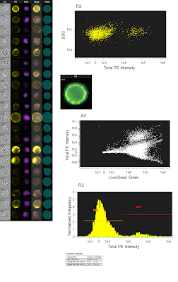
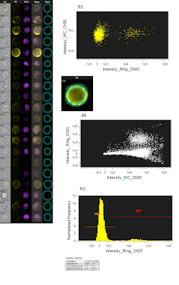 The figure to the left is the originally analyzed data. On the far left is just a gallery of images that show the different fluorescence (the green live/dead wasn't show since dead cells were gated out). The top dot plot is a simple SSC/PE scatter plot to show the distribution of the negative and positives. The image just below is showing the mask used (bluish semitransparent shape overlaying the PE image). Below that is the ungated population showing the Live/Dead Green fluorescence spilling into the PE channel using the whole cell mask. And lastly, a histogram showing the PE Fluorescence. Altogether, a pretty straightforward analysis. However, I wanted to see what would happen if I restricted the PE mask to only the membrane area, so that is what is shown in the figure below. Now, it's important to note that this is the exact same data file, analyzing the exact same group of cells. The only thing changed here is the mask on PE, which is now shown as a ring overlaying the membrane of the cell. If you now look at the SSC/PE scatter plot at the top, you can see the dramatic tightening of the negative population, which implies a reduction of the high autofluorescence cells that were trailing to the right of the negative population in the total cell mask. Another benefit of this restrictive masking strategy was the reduction in the spillover of the green dye into the PE channel as shown in the ungated Live/Dead Green versus PE plot. And lastly, when you look at the histogram, you can see unequivocally the increase in separation between the negatives and positives. To drive the point home a bit more, we can overlay the two histograms so you can see exactly how they match up. Notice that there is a reduction in the intensity of the positive population as well, but this is likely a similar reduction in background fluorescence as is seen in the negative population. The key here and really in all of flow cytometry is RESOLUTION. This is, in fact, what most people are really thinking about when they say 'sensitivity.'
So, can we confirm the original statement here about these imaging cytometers being the most sensitive cytometers available? Well, I'm not sure I'm ready to crown this instrument as the winner just yet, but at least in some circumstances, the ability to only analyze the part of the cell that is actually stained or not stained could prove to be an extremely vital tool especially if you need to resolve dimly stained cells from unstained cells.

It's a really simple example, but in short it involves a surface marker (coupled to PE), a Live/Dead dye (Green) and a Nuclear dye (Violet). By conventional flow cytometry, the PE signal was pretty weak and the user was skeptical that the staining was "real." So, the idea was to make sure the cells were live (Green low/neg), were actually cells (Violet pos) and had surface staining of PE. After going through the normal groups of gating, it came time to look at the PE signal. Surprisingly, it wasn't bad at all (especially with the 561nm laser cranked up to 200mW), however there were some dimmer PE+ cells that were hanging out a bit too close to the negative.
I remember having a discussion with other people in the lab about using carefully calculated masks to pull out the membrane staining and completely removing the cytoplasmic and/or nuclear background which should bring the negative population pretty much down to zero while retaining the specific PE positive fluorescence. This procedure is actually pretty simple so I'll briefly explain it, and if this whole concept of image masking is foreign to you, just think of masks as parts of a cell defined by morphology within which you're going to measure fluorescence. This is very different from flow cytometry where you can only measure fluorescence from the entire cell regardless of where that fluorescence is coming from. With this data, I'm creating a membrane mask, which basically looks like a ring encompassing the outside of the cell. This should retain most of the specific PE fluorescence and remove both background from intracellular autofluorescence, but also background from the nuclear dye and live/dead dye. The figures below demonstrate these findings.

 The figure to the left is the originally analyzed data. On the far left is just a gallery of images that show the different fluorescence (the green live/dead wasn't show since dead cells were gated out). The top dot plot is a simple SSC/PE scatter plot to show the distribution of the negative and positives. The image just below is showing the mask used (bluish semitransparent shape overlaying the PE image). Below that is the ungated population showing the Live/Dead Green fluorescence spilling into the PE channel using the whole cell mask. And lastly, a histogram showing the PE Fluorescence. Altogether, a pretty straightforward analysis. However, I wanted to see what would happen if I restricted the PE mask to only the membrane area, so that is what is shown in the figure below. Now, it's important to note that this is the exact same data file, analyzing the exact same group of cells. The only thing changed here is the mask on PE, which is now shown as a ring overlaying the membrane of the cell. If you now look at the SSC/PE scatter plot at the top, you can see the dramatic tightening of the negative population, which implies a reduction of the high autofluorescence cells that were trailing to the right of the negative population in the total cell mask. Another benefit of this restrictive masking strategy was the reduction in the spillover of the green dye into the PE channel as shown in the ungated Live/Dead Green versus PE plot. And lastly, when you look at the histogram, you can see unequivocally the increase in separation between the negatives and positives. To drive the point home a bit more, we can overlay the two histograms so you can see exactly how they match up. Notice that there is a reduction in the intensity of the positive population as well, but this is likely a similar reduction in background fluorescence as is seen in the negative population. The key here and really in all of flow cytometry is RESOLUTION. This is, in fact, what most people are really thinking about when they say 'sensitivity.'
 So, can we confirm the original statement here about these imaging cytometers being the most sensitive cytometers available? Well, I'm not sure I'm ready to crown this instrument as the winner just yet, but at least in some circumstances, the ability to only analyze the part of the cell that is actually stained or not stained could prove to be an extremely vital tool especially if you need to resolve dimly stained cells from unstained cells.

Thursday, October 13, 2011
Counting Cells with the EMD-Millipore Scepter 2.0
I recently had the chance to play around with the Scepter 2.0 Automatic Cell Counter from EMD-Millipore. The Scepter uses the Coulter Volume principle to count cells in a microfluidic chamber connected to a handheld device. I'm basically using it for things like confirming pre- and post-sort cell counts, as well as counting cells being passaged and primary cells such as PBMCs and splenocytes. The device itself is basically shaped like a pipetteman, and even has a plunger type action which simulates pipetting.
 |
| EMD-Millipore Scepter 2.0 |
 |
| Scepter Software Pro Screenshot |
 |
Blue histogram represents a collection in the upright
position, which leads to low end bubbles showing
up and screwing up the counts. The green histogram
repesents a collection up-side-down and no bubbles
ending up in the chamber leading to way more accurate
counts.
|
Now, about that snafu. What I kept finding was after the sample was loaded into the sensor, and then the sample started traveling through the sensor orifice into the counting micro-channel, I kept seeing bubbles creep in there. The effect of this was I'd start getting these really low volume events piling up near the end of the counting process. It was a small number of low volume events that I could probably gate out (see figure below), but it still messed things up for me. Since I was doing a 1:10 dilution (10ul sample, 90ul buffer), when I back calculate (or better yet, let Scepter Software Pro back-calculate for me) the concentrations, I was off as much as 1x10^6 cells (or a 12% swing in total cell counts). To solve this problem, I made one modification to the collection process. As soon as the sample was loaded into the sensor (it beeps at this point), I immediately flipped the entire Scepter apparatus upside-down as to force any air that begins to enter the sensor to remain near the tip and not enter the orifice and microfluidic channel. This got rid of all the air bubbles and my counts became extremely accurate. In one case, my MoFlo told me there should be 8.02x10^6 cells, and the Scepter counted 8.01x10^6 cells. This made me happy. To see this awesome flip move in action, check out the video below. I apologize for the sound, I was filming this in my sorter room, which has the gentle hum of a twin diesel engine for background noise. Also, you'll just have to trust me when I say "see the bubbles." UPDATE: After playing around with volumes a bit more, it's pretty evident that you definitely need 100+ microliters of volume in your tube. I could get bubbles every time if I only had the requisite 50ul of sample, but if I had 100-120ul, I almost never got bubbles. With this volume, there's no need to turn the scepter upside-down.
So, in all, I think this product was successful for what my purposes were. It's small. The counting process is fast. I can offload the data to my computer, and the counting was very accurate (as long as I remembered to hold it up-side-down to avoid the bubbles). Will I continue to use it? I guess it sort of depends on whether or not I can get over not 'knowing' the %live/dead. For what I'm doing, that's probably fine, but could another option be just as easy and accurate and cheap AND give me live/dead? To be determined. I will say that I've used early versions of the Countess and the Nexcelom, and neither impressed me so much as to make me want to buy one immediately. Hopefully I'll be able to check them out again and perhaps put together a head-to-head review.
Wednesday, October 5, 2011
GLIIFCA 20 Wrap-up.
If you're unfamiliar with the Great Lakes International Imaging and Flow Cytometry Association (GLIIFCA) meeting, you can check out this year's program online here. It's sort of a morph between a technology focused user group meeting and a smaller scale scientific meeting. The focus really is on the utilization of our technology (which I'll refer to under the umbrella term Cytometry) in clinical, translational, and basic research. There is also a strong cytometry vendor presence; about 30 different companies bringing their latest and greatest products. If you'd like to see who attends and supports the association, you can see a list of sponsors on the GLIIFCA site. A part of the meeting that's always a bit disconcerting for me is the Friday night Industrial Science Symposium, which is code-language for "vendor sales pitches." It's been pretty poor some years and not-so-bad others. It really depends on the presentation and the quality of information put forth. You can tell some people are up there literally just trying to sell a product. A good presenter will educate the audience so that the individuals sitting in the chairs come to the conclusion on their own that this is the product they need. And I have to say, we witnessed one of the best examples of this last Friday night in a presentation given by a Chicago-favorite, Kelly Lundsten from BioLegend. Great talk, and actually a pretty good session in total.
The "theme" of the meeting was Cytoinformatics (as opposed to Bioinformatics). As far as the scientific program, it was the first time I found myself thinking, maybe these informatics people aren't wacked. I hear what they're saying, but it usually doesn't strike a chord with me. The basic idea is that you're generating tons of data of various kinds that needs to be quickly integrated in a consistent format in order to support analysis and subsequent decision-making. And I think my resistance has always been in the format of, "Well I don't really generate THAT much data, so I don't have to worry about this stuff." After sitting through a few examples of data generation from some groups that I know pretty well, it got me thinking. The quantity of data can be pretty big even if you're only doing 8-12 parameter flow cytometry or less. This isn't something only for the 18-parameter groups, it's for everyone. Besides the flow data, it would be nice to integrate this info with subject info, imaging info, genomics info, etc.. I think what was pretty successful for this meeting is the fact that it was setup in such a way that you could see the progression of ideas surrounding management of data. 1. Here's the problem: People collect lots of heterogeneous data types. 2. Here's the types of tools needed: Data warehousing, including dimensional models, ETL (extract, transform and load data), and end-user tools to read the relational database. 3. Here are some examples of how people are using these tools with real data and how it impacts decision-making. That was basically GLIIFCA 20, Symposium 1, 2, 3. Kudos to the program committee.
There were also a pretty good crop of posters presented this year, including mine (which won a poster award, thank you very much). Two of them which stuck with me were the "Increased number of laser lines on your cytometer might mess stuff up, so be careful" poster and "Look at this awesome temperature control/antagonist injection apparatus I soldered together with some parts from Home Depot" poster. I'm paraphrasing the titles, of course, and you can find the full poster abstract in the GLIIFCA 20 program linked above. The first one is from the folks just up the road at Northwestern (Geoff Kraker and James Marvin), and the second one comes to us from Roswell Park courtesy of Ed Podniesinski and Paul Wallace. The UCFlow poster was about how "I can't stand looking at QC data, so I'll start using cool Google tools and graphics to make it more interesting and maybe I'll stick with it longer."
So, there you have it. Another year, another GLIIFCA. For the record, this was my 11th GLIIFCA attendance. I have officially attended a majority of GLIIFCA meetings.
 |
| A Slide grabbed from Janet Siebert's (Cytoanalytics) Presentation at GLIIFCA 20 |
 |
| UCFlow's GLIIFCA 20 Poster |
So, there you have it. Another year, another GLIIFCA. For the record, this was my 11th GLIIFCA attendance. I have officially attended a majority of GLIIFCA meetings.
Thursday, September 22, 2011
Safety; It's not to be taken lightly
I'll begin by saying, I definitely need to pay closer attention to the various safety concerns in a lab. All too often we sacrifice our own safety in order to get things done quicker; cutting corners, thinking I'll be careful. And then, bam, you have an incident that you regret. Fortunately, I haven't had to deal with this first hand, but what I'm going to describe here happened close enough to home that it caused me to pause for a minute and evaluate my own techniques and protocols in the lab.
Perhaps some of you are aware of a recent incident at the University of Chicago, where a scientist became infected with the same strain of bacteria that is being studied in the lab (B. cereus). According to information published on the Science Magazine site, the infected individual was not even working with the microbe but may have transfered it to an uncovered wound via a spill (http://news.sciencemag.org/scienceinsider/2011/09/university-of-chicago-microbiologist.html). I believe the infected person is going to be fine and needed to undergo surgery to remove the infected tissue, so that's positive. As a result of this (and another incident just 2 years ago), the PI is moving these sets of experiments to Argonne National Labs in the Howard T. Ricketts lab, where they are also running experiments on Plague, MRSA, and Anthrax.
It was roughly two years ago to the day that a researcher in this same Laboratory at the University of Chicago died from exposure to an attenuated form of Y. pestis. In this case, the researcher may have felt a bit safer than he was, since the strain was determined to be non-lethal. His co-workers admitted that his glove wearing practices were inconsistent at best. It just so happened that he also had an undetected/untreated condition known as hemochromatosis, or an overload of iron in the blood. It may have been this overload of iron that allowed the attenuated version of the bacteria to become virulent (http://en.wikipedia.org/wiki/Malcolm_Casadaban).
So, as you can see, we have plenty of examples of the potential threat to our safety and those around us, and we should use examples like these, not to place blame on those who made mistakes, but to remind us of the importance to slow down and think about what we're doing and what we need to do to stay safe. There's really just two reasons why incidents like this happen; Carelessness or Ignorance. You have to be aware of what you're working with. Ask questions if you're unsure. Educate yourself. Nothing is so important that you cannot take the extra steps to make sure you and those around you are protected as much as possible.
Those who work in your safety office are not out to get you. They're here to educate first and foremost, and yes, to enforce standard operating procedures for your protection. In perusing our own safety department's web site, I stumbled upon this - Shared Responsibility.
Mission: http://safety.uchicago.edu/about/index.shtml
Environmental Health and Safety provides services and support for efficient, effective, and compliant work practices, while promoting a culture of shared responsibility by students, faculty, staff and visitors for a healthy, safe, and environmentally sound educational and research community at the University of Chicago.
So, hopefully if you've taken the time to read through this, you can certainly take the time to re-evaluate the procedures in your lab. Make a plan to educated your staff and those around you, and open the lines of communication between your lab and those who have been tasked with the safety of your institution. Stay safe!
Perhaps some of you are aware of a recent incident at the University of Chicago, where a scientist became infected with the same strain of bacteria that is being studied in the lab (B. cereus). According to information published on the Science Magazine site, the infected individual was not even working with the microbe but may have transfered it to an uncovered wound via a spill (http://news.sciencemag.org/scienceinsider/2011/09/university-of-chicago-microbiologist.html). I believe the infected person is going to be fine and needed to undergo surgery to remove the infected tissue, so that's positive. As a result of this (and another incident just 2 years ago), the PI is moving these sets of experiments to Argonne National Labs in the Howard T. Ricketts lab, where they are also running experiments on Plague, MRSA, and Anthrax.
It was roughly two years ago to the day that a researcher in this same Laboratory at the University of Chicago died from exposure to an attenuated form of Y. pestis. In this case, the researcher may have felt a bit safer than he was, since the strain was determined to be non-lethal. His co-workers admitted that his glove wearing practices were inconsistent at best. It just so happened that he also had an undetected/untreated condition known as hemochromatosis, or an overload of iron in the blood. It may have been this overload of iron that allowed the attenuated version of the bacteria to become virulent (http://en.wikipedia.org/wiki/Malcolm_Casadaban).
So, as you can see, we have plenty of examples of the potential threat to our safety and those around us, and we should use examples like these, not to place blame on those who made mistakes, but to remind us of the importance to slow down and think about what we're doing and what we need to do to stay safe. There's really just two reasons why incidents like this happen; Carelessness or Ignorance. You have to be aware of what you're working with. Ask questions if you're unsure. Educate yourself. Nothing is so important that you cannot take the extra steps to make sure you and those around you are protected as much as possible.
Those who work in your safety office are not out to get you. They're here to educate first and foremost, and yes, to enforce standard operating procedures for your protection. In perusing our own safety department's web site, I stumbled upon this - Shared Responsibility.
Mission: http://safety.uchicago.edu/about/index.shtml
Environmental Health and Safety provides services and support for efficient, effective, and compliant work practices, while promoting a culture of shared responsibility by students, faculty, staff and visitors for a healthy, safe, and environmentally sound educational and research community at the University of Chicago.
And specifically regarding Laboratory Safety, they have this to add: http://safety.uchicago.edu/labpersonnel/index.shtml
Research is one of the two main missions of the University, the other being education. Lab personnel are integral in the creation and maintenance of a safe laboratory environment. They are responsible for ensuring safety in laboratories and lab support areas on campus and within the Medical Center. This responsibility includes:
- Being familiar with University emergency procedures;
- Responding appropriately in the event of an emergency;
- Being familiar with Environmental Health and Safety policies and procedures;
- Maintaining a safe laboratory environment;
- Knowing the hazards of the materials and/or equipment being used;
- Following all safety procedures in the laboratory environment;
- Selecting, using and understanding the limitations of personal protective equipment;
- Reporting any unsafe conditions to your supervisor and/or Environmental Health and Safety; and
- Reporting any job related injuries or illnesses to your supervisor or Human Resources Administrator immediately; and
- Participating in all required safety training.
So, hopefully if you've taken the time to read through this, you can certainly take the time to re-evaluate the procedures in your lab. Make a plan to educated your staff and those around you, and open the lines of communication between your lab and those who have been tasked with the safety of your institution. Stay safe!
Subscribe to:
Posts (Atom)